Fraud Blocker Skip to main content

Capillary Collection (page 7)

"Exploring the Intricate Network of Capillaries: A Journey through the Human Digestive System and Artistic Renderings" Step into the mesmerizing world of capillaries

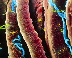
Background imageCapillary Collection: False-colour SEM of skeletal muscle fibres

False-colour SEM of skeletal muscle fibres
Muscle. False-colour scanning electron micrograph of a bundle of skeletal muscle fibres (red), also known as striated muscle fibres, and a few capillaries (blue)

Background imageCapillary Collection: Fracture, computer artwork

Fracture, computer artwork
Bone fracture. Computer artwork showing detail of a bone fracture. The capillary (red) has remained intact. All fractures are reset into position and the bone heals naturally

Background imageCapillary Collection: Kidney disease, TEM

Kidney disease, TEM
Kidney disease. Coloured transmission electron micrograph (TEM) of a section through the glomerulus of a kidney affected by membranous neuropathy

Background imageCapillary Collection: Marcello Malpighi, Italian biologist

Marcello Malpighi, Italian biologist
Marcello Malpighi (1628-1694), Italian biologist. Malpighi was a physician by training, graduating at Bologna in 1653. Malpighis observations through his microscope were to revolutionise anatomy

Background imageCapillary Collection: Inflamed kidney, light micrograph

Inflamed kidney, light micrograph
Inflamed kidney. Light micrograph of a section through a kidney with acute nephritis (inflammation). There are areas of necrosis (tissue death) within the glomerulus (centre)

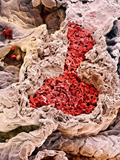
Background imageCapillary Collection: Lung blood vessel, SEM

Lung blood vessel, SEM
Lung blood vessel. Coloured scanning electron micrograph (SEM) of blood vessels around alveoli (air spaces, dark brown) in the lungs. Erythrocytes (red blood cells) are seen in the blood vessel

Background imageCapillary Collection: Arteries, computer artwork

Arteries, computer artwork

Background imageCapillary Collection: Skin blood supply, artwork

Skin blood supply, artwork
Skin blood supply. Computer artwork showing the structure of the microscopic loops of blood vessels that supply the epidermis, the outer layer of the skin

Background imageCapillary Collection: Glomerular capillary, TEM

Glomerular capillary, TEM
Glomerular capillary. Coloured transmission electron micrograph (TEM) of a section through kidney tissue, showing a capillary from a glomerulus. Glomeruli filter waste products from the blood

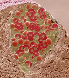
Background imageCapillary Collection: Retina blood vessel, SEM

Retina blood vessel, SEM
Retina blood vessel. Coloured scanning electron micrograph (SEM) of a section through a blood vessel in the retina of an eye, showing the red blood cells (red)

Background imageCapillary Collection: Lung alveolus blood supply, artwork

Lung alveolus blood supply, artwork. The alveoli (air sacs) are the site of gaseous exchange in the lungs. Carbon dioxide from de-oxygenated red blood cells (blue) passes from the capillaries

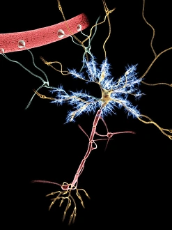
Background imageCapillary Collection: Nerve cells, artwork

Nerve cells, artwork
Neuron, nerve cell, firing, computer artwork

Background imageCapillary Collection: Blood vessel, artwork

Blood vessel, artwork
Blood vessel. Computer artwork of the inside of a blood vessel, showing red blood cells (circular) and the epithelial cells linging the vessels wall

Background imageCapillary Collection: Retina blood vessels and nerve cells

Retina blood vessels and nerve cells. Light micrograph showing the blood vessels and nerve cells in the retina of an eye. The retina is the light-sensitive membrane that lines the back of the eyeball

Background imageCapillary Collection: Capillary, SEM

Capillary, SEM
Capillary. Coloured scanning electron micrograph (SEM) of a red blood cell (erythrocyte) in a capillary. Magnification: x9000 when printed at 10 centimetres tall

Background imageCapillary Collection: Small intestine structures, artwork

Small intestine structures, artwork
Small intestine structures. Artwork sequence of magnified views of the small intestine, with the magnification increasing from top to bottom

Background imageCapillary Collection: Intestinal villi anatomy, artwork

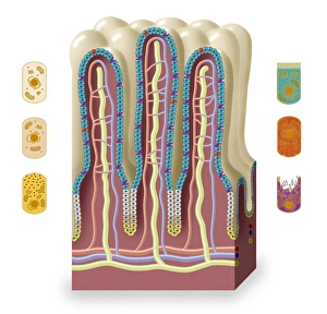
Intestinal villi anatomy, artwork
Intestinal villi anatomy. Artwork showing six types of cell found on the surface of villi in the small intestine. Clockwise from upper right they are: enterocytes (green)

Background imageCapillary Collection: Bowmans capsule, diagram

Bowmans capsule, diagram
Bowmans capsule. Diagram showing the anatomical structure of a Bowmans capsule and glomerulus in a kidney

Background imageCapillary Collection: Lung alveolus, artwork

Lung alveolus, artwork
Lung alveolus, computer artwork. Alveoli are the site of gas exchange in the lungs of mammals, where oxygen is taken up and carbon dioxide released

Background imageCapillary Collection: Capillary, artwork

Capillary, artwork
Capillary. Artwork showing the anatomical structure of a capillary seen in cross-section

Background imageCapillary Collection: Unmyelinated nerve bundle, TEM

Unmyelinated nerve bundle, TEM
Unmyelinated nerve bundle. Coloured transmission electron micrograph (TEM) of a section through a group of unmylinated nerve fibres

Background imageCapillary Collection: Migraine mechanisms, artwork

Migraine mechanisms, artwork
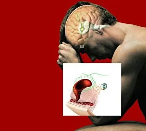
Migraine mechanisms. Artwork showing the blood vessel and brain mechanisms thought to cause some types of migraines. It is thought that the trigger is nitric oxide (NO)

Background imageCapillary Collection: Retina in glaucoma, artwork

Retina in glaucoma, artwork
Retina in glaucoma, computer artwork. This view is looking through the pupil (green ring) at the front of the eye, to the retina (orange) at the back of the eye

Background imageCapillary Collection: Kidney glomerulus, light micrograph

Kidney glomerulus, light micrograph
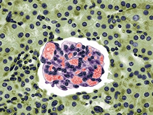
Kidney glomerulus. Coloured light micrograph of a section through the cortex of a healthy kidney, showing a glomerulus. The core of a glomerulus is formed of a tightly coiled capillary (purple)

Background imageCapillary Collection: Intestinal blood vessels, SEM

Intestinal blood vessels, SEM
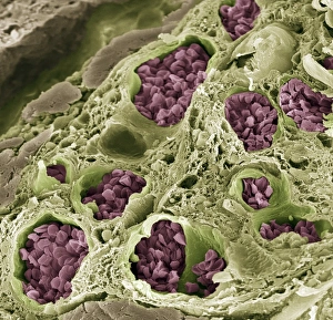
Intestinal blood vessels. Coloured scanning electron micrograph (SEM) of a freeze fracture section through the lining of the colon

Background imageCapillary Collection: Skin tissue, light micrograph

Skin tissue, light micrograph
Skin tissue. Light micrograph of a transverse section through human skin. The skin is made up of an outer epidermis (pale purple, across top) with hair follicles (deep purple)

Background imageCapillary Collection: Dogfish gill, light micrograph

Dogfish gill, light micrograph
Dogfish gill. Light micrograph of a section through a gill lamella from a lesser spotted dogfish (Scyliorhinus canicula). Running across centre is a brachial artery

Background imageCapillary Collection: Thyroid follicle structures, TEM

Thyroid follicle structures, TEM
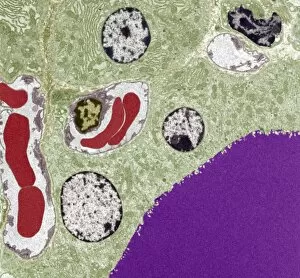
Thyroid follicle structures. Coloured transmission electron micrograph (TEM) of a section through structures in a thyroid follicle
Background imageCapillary Collection: Foot skin tissue, light micrograph

Foot skin tissue, light micrograph
Foot skin tissue. Light micrograph of a transverse section through skin from a human foot. The skin is made up of an outer epidermis (across top)


All Professionally Made to Order for Quick Shipping

"Exploring the Intricate Network of Capillaries: A Journey through the Human Digestive System and Artistic Renderings" Step into the mesmerizing world of capillaries, where intricate pathways weave their way through our bodies. In this captivating journey, we delve into various organs and tissues to uncover the beauty and functionality of these tiny blood vessels. Starting with a closer look at the kidney glomerulus under a scanning electron microscope (SEM), we witness its complex structure that filters waste products from our blood. The delicate web-like arrangement of capillaries ensures efficient filtration, enabling our kidneys to maintain proper balance in our body. Moving on to another SEM image, we discover thyroid gland capillaries intricately intertwined within this vital endocrine organ. These microscopic vessels play a crucial role in delivering hormones throughout our body, ensuring proper metabolism and growth. As we continue exploring kidney glomeruli under SEM once again, their unique architecture comes into focus. These clusters of capillaries enable selective reabsorption and excretion processes necessary for maintaining fluid balance in our bodies. Shifting gears slightly, we turn towards retina blood vessels captured by SEM. Here, these tiny capillaries supply oxygen-rich blood to nourish the light-sensitive cells responsible for vision – an exquisite example of nature's design meeting function. Intriguingly, computer artwork showcases red blood cells flowing gracefully through capillary networks. This imaginative representation reminds us how essential these vessels are in transporting oxygen and nutrients while removing waste products throughout every corner of our bodies. Diving deeper into muscle tissue with false-color SEM imagery reveals pericyte-covered capillaries. Pericytes act as guardians protecting these fragile vessels while also playing a role in regulating blood flow – an elegant symbiotic relationship between cells within us. Concluding this captivating journey is yet another glimpse at kidney glomeruli but now observed through light microscopy. This traditional technique highlights their distinct appearance and allows us to appreciate the intricate details of these vital structures.