
Blood Clot Collection
"Exploring the Intricate World of Blood Clots: Unveiling their Formation and Impact" SEM C016 / 9747


All Professionally Made to Order for Quick Shipping
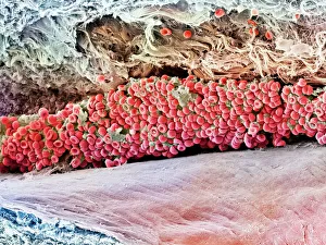
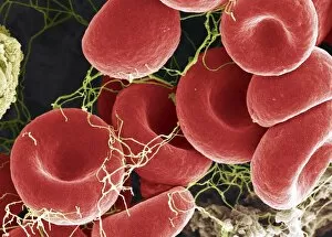
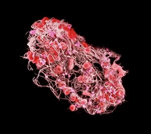
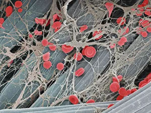
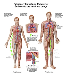
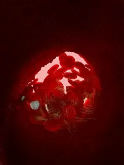
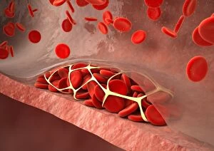
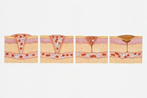
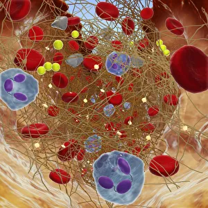
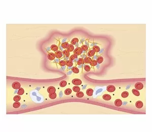
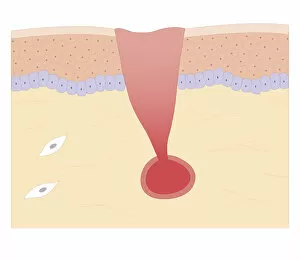
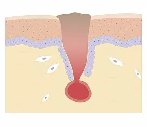
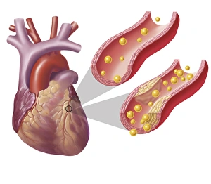
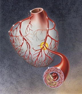
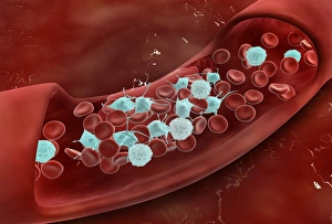
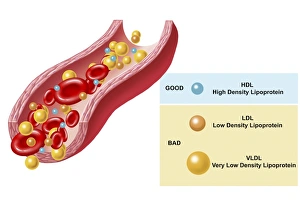
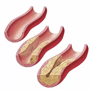
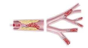
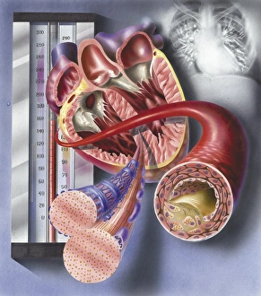
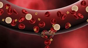
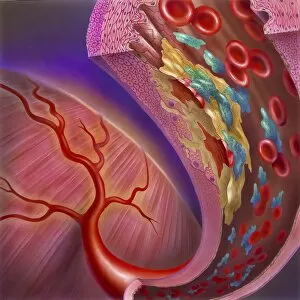
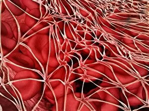
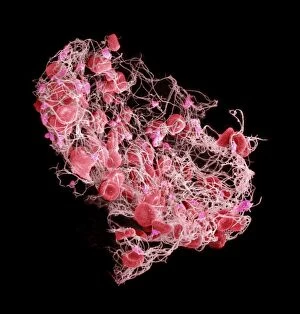
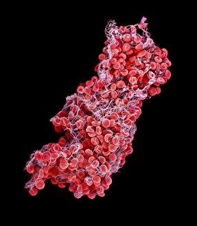
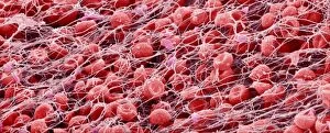
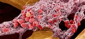
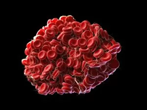
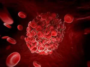
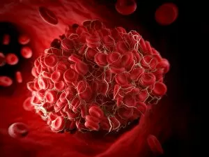
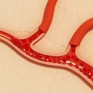
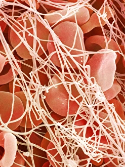
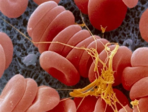
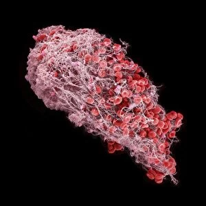
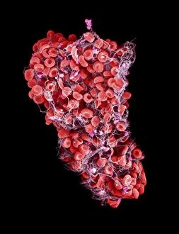
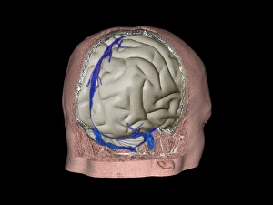
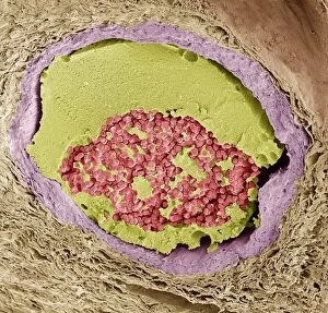
"Exploring the Intricate World of Blood Clots: Unveiling their Formation and Impact" SEM C016 / 9747: Witness the mesmerizing beauty of a scanning electron micrograph (SEM) capturing a red blood cell amidst a blood clot, showcasing its intricate structure. Blood clot on plaster, SEM: Delve into the microscopic realm as we observe a detailed view of a blood clot formed on a plaster surface through an SEM image. Unraveling the Mystery: A captivating SEM image reveals the complexity of a blood clot, shedding light on its composition and formation process. Pulmonary Embolism Pathway Revealed: Explore how an embolus travels to reach the heart and lungs in this informative depiction of pulmonary embolism's path. Artwork C016 / 4619: Discover an artistic representation that portrays the essence of a blood clot, merging science with creativity for visual impact. White Blood Cells and Platelets Under Magnification: Observe white blood cells and platelets magnified in an SEM image (C016 / 3099), offering insight into their role within blood clots. The Dynamic Duo at Work: Dive deeper into another captivating SEM image (C016 / 3098) revealing white blood cells and platelets collaborating to form essential components of blood clots. Thrombosed Blood Vessel Artwork Explored: Immerse yourself in an artwork (C013 / 4649) illustrating thrombosis—a condition where damaged vessels constrict, leading to potentially dangerous clots forming inside them. Step-by-Step Insight Into Clot Formation Illustrated: Engage with four cross-sectional illustrations depicting how platelet plugs form by tracing the journey from oozing wound to complete coagulation—educational yet visually striking. Cross Section Biomedical Illustration Exposed.