
Biotechnology Collection (page 2)
"Unveiling the Future: Biotechnology Revolutionizing Our World" Step into a world where boundaries blur and possibilities expand as biotechnology takes center stage


All Professionally Made to Order for Quick Shipping
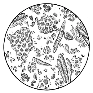
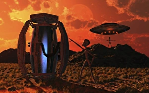
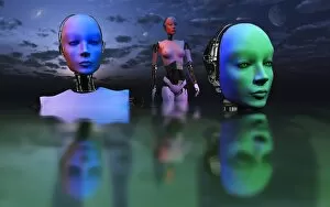
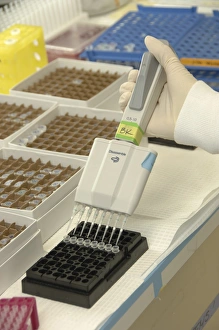
"Unveiling the Future: Biotechnology Revolutionizing Our World" Step into a world where boundaries blur and possibilities expand as biotechnology takes center stage. In this captivating image, a transparent man walks through the vast expanse of a desert, symbolizing the limitless potential that lies ahead. Picture No. 12479759 showcases cutting-edge advancements in artificial intelligence (AI), androids, and augmented reality (AR). With arms raised in triumph, these technological marvels redefine what it means to be human. Gone are the days of back pain and limitations; bionic enhancements offer us an unprecedented level of freedom and capability. Delving deeper into history, Picture No. 12479758 transports us to Washington D. C. , circa 1926. Witnessing scientists huddled around flasks brimming with promise, we glimpse the birth of biotechnological breakthroughs that would shape our modern world. In another frame, we find ourselves peering through the lens of scientific discovery as a dedicated scientist gazes intently into a microscope. The science laboratory buzzes with excitement as researchers unravel mysteries at microscopic levels – unlocking secrets that will revolutionize medicine and beyond. A person engrossed in their own microscopic exploration reminds us that curiosity fuels progress. Through relentless pursuit of knowledge, we uncover groundbreaking solutions to complex challenges - from curing diseases to improving agricultural practices. But it's not just humans pushing boundaries; even robots join this transformative journey towards advancement. A robot woman weighs in on matters both physical and metaphorical – representing how automation can enhance efficiency while also sparking debates about ethics and humanity's role in an increasingly automated world. As acetylene illuminates dark corners within laboratories equipped with state-of-the-art technology, AI-driven systems work tirelessly alongside passionate scientists who strive for excellence amidst adversity. This fusion between advanced technology and human ingenuity propels us forward towards unimaginable achievements. Biotechnology is more than just science; it's a testament to our unwavering affection for progress.