Fraud Blocker Skip to main content

Biochemical Collection (page 6)

"Unveiling the Intricacies of Biochemical World: From Anaesthetic Inhibiting Ion Channels to DNA Discoveries" Delving into the depths wonders

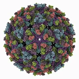
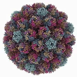
Background imageBiochemical Collection: Chikungunya virus capsid

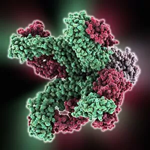
Chikungunya virus capsid, molecular model. This virus, transmitted by mosquitoes in tropical Africa and Asia, causes fever and joint pain in humans, similar to dengue fever

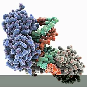
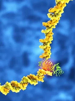
Background imageBiochemical Collection: TATA box-binding protein complex C014 / 0867

TATA box-binding protein complex C014 / 0867
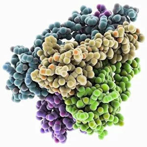
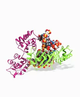
TATA box-binding protein complex. Molecular model showing a TATA box-binding protein (TBP, khaki) complexed with a strand of DNA (deoxyribonucleic acid)

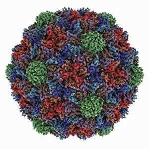
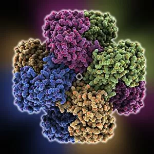
Background imageBiochemical Collection: HK97 bacteriophage procapsid

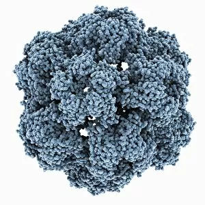
HK97 bacteriophage procapsid. Molecular model showing the structure of the prohead-I procapsid of the HK97 bacteriophage. Bacteriophages are viruses that infect bacteria

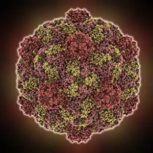
Background imageBiochemical Collection: Turnip yellow mosaic virus capsid

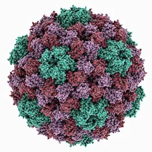
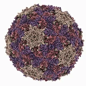
Turnip yellow mosaic virus capsid, molecular model. This virus infects a wide variety of plants, including crops such as turnips and cabbages, causing yellow patches on the leaves

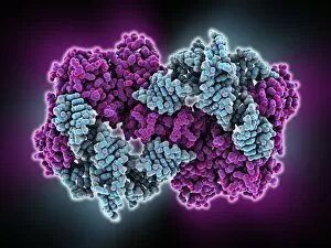
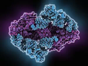
Background imageBiochemical Collection: E. coli Holliday junction complex C014 / 0878

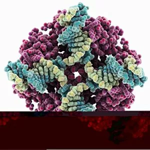
E. coli Holliday junction complex C014 / 0878
E. coli Holliday junction complex. Molecular model of a RuvA protein (dark pink) in complex with a Holliday junction between homologous strands of DNA (deoxyribonucleic acid)

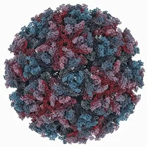
Background imageBiochemical Collection: Sindbis virus capsid, molecular model

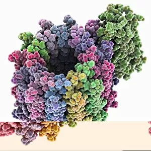
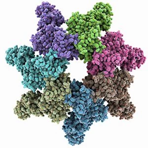
Sindbis virus capsid, molecular model. This virus, transmitted by mosquitoes, causes sindbis fever in humans. In viruses, the capsid is the protein shell that encloses the genetic material

Background imageBiochemical Collection: Fibrinogen molecule C014 / 0473

Fibrinogen molecule C014 / 0473
Fibrinogen. Molecular model showing the structure of the blood clotting glycoprotein fibrinogen (factor I). The molecule consists of two sets of alpha (grey)

Background imageBiochemical Collection: Genetic research, conceptual image C014 / 1256

Genetic research, conceptual image C014 / 1256
Genetic research. Conceptual image of a molecular model of a strand of DNA (deoxyribonucleic acid) being held on a human hand

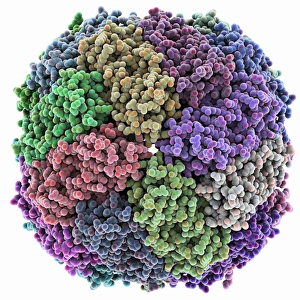
Background imageBiochemical Collection: Murine polyomavirus capsid

Murine polyomavirus capsid, molecular model. This virus, one of a range named for their potential to cause multiple tumours, infects mice

Background imageBiochemical Collection: Human antitumour antibody molecule

Human antitumour antibody molecule
Human antitumour antibody. Molecular model showing the antitumour antibody BR96 complexed with part of the Lewis antigen. The Lewis antigen is expressed on the surface of human carcinoma cells

Background imageBiochemical Collection: Iron containing protein, molecular model

Iron containing protein, molecular model
Iron containing protein. Molecular model showing the structure of a bacterial homolog of the animal iron containing protein ferritin

Background imageBiochemical Collection: Brome mosaic virus capsid

Brome mosaic virus capsid, molecular model. This plant virus infects grasses, especially brome grasses, and also barley. It causes mosaic patches of discolouration

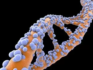
Background imageBiochemical Collection: Synthetic DNA molecule

Synthetic DNA molecule
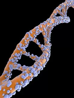
Synthetic DNA. Molecule model of a synthetic form of DNA (deoxyribonucleic acid). DNA is composed of two strands twisted into a double helix

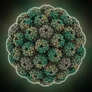
Background imageBiochemical Collection: Cowpea chlorotic mottle virus capsid

Cowpea chlorotic mottle virus capsid, molecular model. This virus (CCMV) infects the cowpea plant (Vigna unguiculata), causing yellow spots of discolouration

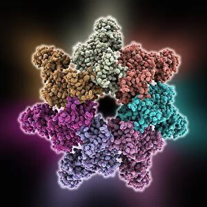
Background imageBiochemical Collection: Potassium ion channel protein structure

Potassium ion channel protein structure. Molecular model of a KcsA potassium ion (K+) channel from Streptomyces lividans bacteria

Background imageBiochemical Collection: Streptavidin-biotin molecular complex

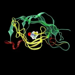
Streptavidin-biotin molecular complex. Molecular model of a single-strand binding complex of streptavidin (ribbons) and biotin (space-filled model, centre). Biotin is also known as vitamin B7

Background imageBiochemical Collection: DNA supercoil, artwork

DNA supercoil, artwork
DNA supercoils. Computer artwork showing a supercoiled strand of DNA (deoxyribonucleic acid). Supercoiling is important in a number of biological processes

Background imageBiochemical Collection: Potassium ion channel beta subunit

Potassium ion channel beta subunit. Molecular model showing the structure a beta subunit of a voltage-dependent potassium (K+) channel

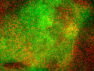
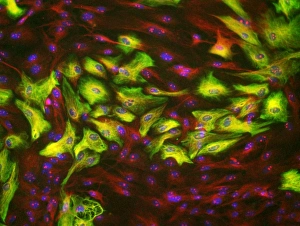
Background imageBiochemical Collection: Stem cell-derived retinal cells

Stem cell-derived retinal cells. Fluorescence light micrograph of retinal pigment epithelium (RPE) cells that have been derived from human embryonic stem cells (HESC)

Background imageBiochemical Collection: Tyrosyl-tRNA synthetase molecule

Tyrosyl-tRNA synthetase molecule
Tyrosyl-tRNA synthetase protein molecule. Molecular model showing bacterial tyrosyl-tRNA synthetase complexed with tyrosyl tRNA (transfer ribonucleic acid)

Background imageBiochemical Collection: KCNQ ion channel protein structure

KCNQ ion channel protein structure. Molecular model showing the protein structure of an ion channel domain. Ion channels are membrane-spanning proteins that form a pathway for the movement of

Background imageBiochemical Collection: Potassium ion channel cavity structure

Potassium ion channel cavity structure. Molecular model showing the structure of a cavity formed by potassium ion channel proteins

Background imageBiochemical Collection: Stem cell-derived neurons, micrograph

Stem cell-derived neurons, micrograph
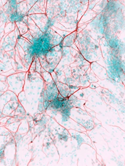
Stem cell-derived neurons. Light micrograph of human nerve cells (neurons) that have been derived from induced pluripotent stem cells (IPS). Tuj1 proteins are cyan, and cell nuclei are red

Background imageBiochemical Collection: Endonuclease IV molecule

Endonuclease IV molecule. Molecular model of the endonuclease IV restriction enzyme EcoRV (grey) bound to a cleaved section of DNA (deoxyribonucleic acid, blue, orange and pink)

Background imageBiochemical Collection: Tumour suppressor protein and DNA C017 / 3645

Tumour suppressor protein and DNA C017 / 3645
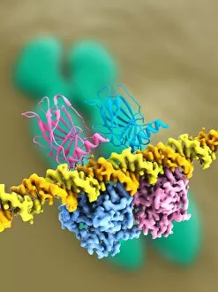
Tumour suppressor protein and DNA. Computer artwork showing a molecule of the tumour suppressor protein p53 (blue and pink) bound to a molecule of DNA (deoxyribonucleic acid, yellow and orange)

Background imageBiochemical Collection: ATP synthase molecule C014 / 0880

ATP synthase molecule C014 / 0880
ATP synthase molecule. Molecular model showing the structure of ATP synthase (ATPase) subunit C. ATPase is an important enzyme that provides energy for cells through the synthesis of adenosine

Background imageBiochemical Collection: Avian polyomavirus capsid

Avian polyomavirus capsid, molecular model. This virus, one of a range named for their potential to cause multiple tumours, infects birds. Discovered in budgerigars in 1981, it is often fatal

Background imageBiochemical Collection: Astrocyte brain cells, light micrograph

Astrocyte brain cells, light micrograph
Astrocyte brain cells. Fluorescence light micrograph of primary astrocyte cells from the brain of a mouse. Astrocytes have numerous branches of connective tissue that provide support

Background imageBiochemical Collection: DNA 6-way junction, artwork C014 / 2587

DNA 6-way junction, artwork C014 / 2587
DNA 6-way junction. Computer artwork of a synthetic assemblage of nucleic acids which are useful in the design of nanostructures

Background imageBiochemical Collection: Anthrax protective antigen molecule C014 / 0886

Anthrax protective antigen molecule C014 / 0886
Anthrax protective antigen molecule. Computer model showing the structure of a molecule of protective antigen (PA) produced by anthrax (Bacillus anthracis) bacteria

Background imageBiochemical Collection: HIV enzyme protein, molecular model C014 / 0876

HIV enzyme protein, molecular model C014 / 0876
HIV enzyme protein. Computer model showing the structure of the catalytic domain of a molecule of HIV-1 retroviral integrase (IN) from the human immunodeficiency virus (HIV)

Background imageBiochemical Collection: Tryptophanyl-tRNA synthetase molecule

Tryptophanyl-tRNA synthetase molecule
Tryptophanyl-tRNA synthetase protein molecule. Molecular model showing human tryptophanyl-tRNA synthetase complexed with tryptophan tRNA (transfer ribonucleic acid)

Background imageBiochemical Collection: Cytoplasmic polyhedrosis virus capsid

Cytoplasmic polyhedrosis virus capsid, molecular model. Part of the Cypovirus genus and invariably fatal, this insect virus is transmitted by contamination of leaves eaten (examples include silkworms)

Background imageBiochemical Collection: EcoRV restriction enzyme molecule C014 / 2117

EcoRV restriction enzyme molecule C014 / 2117
EcoRV restriction enzyme. Molecular model of the type II restriction enzyme EcoRV (purple and blue) bound to a DNA molecule (deoxyribonucleic acid, pink and white)

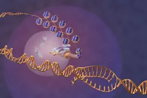
Background imageBiochemical Collection: Gene expression, artwork

Gene expression, artwork
Gene expression. Computer artwork showing the process of transcription, the first stage or gene expression. Here, a chromosome (distance)

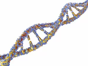
Background imageBiochemical Collection: DNA molecule, artwork F007 / 1996

DNA molecule, artwork F007 / 1996
DNA molecule, computer artwork

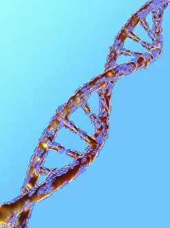
Background imageBiochemical Collection: DNA molecule, artwork F007 / 1994

DNA molecule, artwork F007 / 1994
DNA molecule, computer artwork

Background imageBiochemical Collection: DNA molecule, artwork F007 / 1995

DNA molecule, artwork F007 / 1995
DNA molecule, computer artwork

Background imageBiochemical Collection: DNA molecule, artwork F007 / 1991

DNA molecule, artwork F007 / 1991
DNA molecule, computer artwork

Background imageBiochemical Collection: DNA molecule, artwork F007 / 1992

DNA molecule, artwork F007 / 1992
DNA molecule, computer artwork

Background imageBiochemical Collection: HIV enzyme protein, molecular model

HIV enzyme protein, molecular model
HIV enzyme protein. Computer model showing the structure of the catalytic domain of a molecule of HIV-1 retroviral integrase (IN) from the human immunodeficiency virus (HIV)

Background imageBiochemical Collection: Anthrax protective antigen molecule C014 / 0865

Anthrax protective antigen molecule C014 / 0865
Anthrax protective antigen molecule. Computer model showing the structure of a molecule of protective antigen (PA) produced by anthrax (Bacillus anthracis) bacteria

Background imageBiochemical Collection: EcoRV restriction enzyme molecule C014 / 2112

EcoRV restriction enzyme molecule C014 / 2112
EcoRV restriction enzyme. Molecular model of the type II restriction enzyme EcoRV (pink) bound to a cleaved section of DNA (deoxyribonucleic acid, yellow)

Background imageBiochemical Collection: TATA box-binding protein complex C017 / 7090

TATA box-binding protein complex C017 / 7090
TATA box-binding protein complex. Molecular model showing a TATA box-binding protein (TBP, green) complexed with a strand of DNA (deoxyribonucleic acid, spheres) and transcription factor IIB

Background imageBiochemical Collection: Theilers encephalomyelitis virus capsid

Theilers encephalomyelitis virus capsid, molecular model. This virus, which causes brain and spinal cord inflammation in mice, is used in research

Background imageBiochemical Collection: TATA box-binding protein complex C017 / 7085

TATA box-binding protein complex C017 / 7085
TATA box-binding protein complex. Molecular model showing a TATA box-binding protein (TBP, green) complexed with a strand of DNA (deoxyribonucleic acid, yellow) and transcription factor IIB

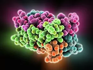
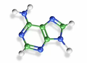
Background imageBiochemical Collection: Adenine molecule, artwork C017 / 7199

Adenine molecule, artwork C017 / 7199
Adenine molecule. Computer artwork showing the structure of a molecule of the nucleobase adenine. Atoms are colour-coded spheres: carbon (green), nitrogen (blue), and oxygen (white)

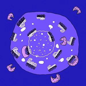
Background imageBiochemical Collection: Wnt signalling pathways, illustration C018 / 0917

Wnt signalling pathways, illustration C018 / 0917
Wnt signalling pathways, illustration. Wnt signalling pathways are three separate pathways that pass signals from outside a cell to inside the cell


All Professionally Made to Order for Quick Shipping

"Unveiling the Intricacies of Biochemical World: From Anaesthetic Inhibiting Ion Channels to DNA Discoveries" Delving into the depths wonders, scientists have uncovered an anaesthetic that inhibits an ion channel (C015 / 6718), shedding light on new possibilities for pain management. The enigmatic double-stranded RNA molecule reveals its secrets, captivating researchers with its role in gene regulation and potential therapeutic applications. Peering into the intricate world of DNA transcription through a molecular model, scientists unravel the mysteries behind genetic information transfer and cellular function. Captured under a microscope's gaze, caffeine crystals dazzle with their vibrant beauty, reminding us of this ubiquitous stimulant's impact on our daily lives. The iconic DNA molecule stands tall as a symbol of life's blueprint, holding within it the key to our genetic heritage and evolutionary history. Immunoglobulin G antibody molecule emerges as a formidable defender against pathogens, showcasing nature's ingenious immune system at work. Through mesmerizing crystal formations seen under intense magnification, EDTA crystals reveal their significance in chelation therapy and metal ion sequestration processes. Oxytocin hormone crystals shimmer like precious gems when observed through polarized light microscopy (PLM C016 / 7196), highlighting its crucial role in social bonding and reproductive functions. Watson and Crick forever etched their names in scientific history by unravelling the structure of DNA; their groundbreaking discovery paved the way for countless advancements in genetics research. Celebrated author Isaac Asimov not only captivated readers with his science fiction tales but also left an indelible mark as a biochemist who popularized complex scientific concepts for all to comprehend and appreciate. Artistic renditions bring metabolic enzymes to life as they orchestrate vital chemical reactions within cells – true catalysts that drive life's intricate processes.