
Beta Sheets Collection
"Unraveling the Beauty of Beta Sheets: A Molecular Masterpiece" In the intricate world of molecular biology


All Professionally Made to Order for Quick Shipping
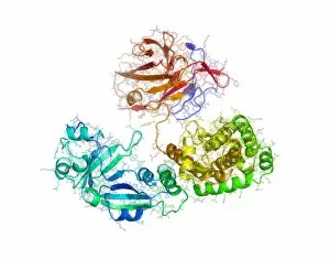
"Unraveling the Beauty of Beta Sheets: A Molecular Masterpiece" In the intricate world of molecular biology, beta sheets stand as captivating structures that have revolutionized our understanding of various biological processes. From Interleukin-6 to Insulin receptor substrate 1 molecule, these fascinating entities have left an indelible mark on scientific research. Nobel Prize-worthy in their significance, they can not just mere conceptual artwork but rather a fundamental component of proteins. They play a crucial role in maintaining protein stability and function by forming hydrogen bonds between adjacent strands. Delving deeper into their versatility they can be found in diverse molecules such as Adiponectin hormone molecule and Cholinesterase enzyme. Their presence is even evident in notorious viruses like the Protein from outer coat of SARS virus and Influenza A virus haemagglutinin protein. Beta sheets continue to captivate researchers with their involvement in deadly pathogens like Anthrax lethal factor protein and FMD virus surface protein. These structures hold vital clues for developing effective treatments against infectious diseases. The elegance extends beyond viral realms; they also contribute to essential enzymes like Reverse transcriptase enzyme from HIV and Influenza B virus neuraminidase enzyme. Understanding their structure aids scientists in designing targeted therapies against these formidable foes. One cannot overlook the aesthetic appeal when observing Bluetongue virus protein VP7 structure - a mesmerizing example showcasing the intricacy hidden within nature's building blocks. As we unravel the secrets held within these remarkable formations, we inch closer towards unlocking breakthroughs that could shape medical advancements for generations to come. Beta sheets serve as both artistic masterpieces and scientific wonders, reminding us that beauty lies not only on canvas but also within the microscopic realm where life's mysteries unfold.