Fraud Blocker Skip to main content

Bacteria Collection (page 4)

"Bacteria: The Microscopic Marvels of Yellowstone National Park" In the heart of Wyoming, USA lies the breathtaking Yellowstone National Park

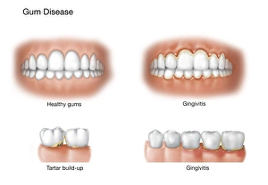
Background imageBacteria Collection: Comparison of healthy gums versus gingivitis

Comparison of healthy gums versus gingivitis

Background imageBacteria Collection: Picture No. 11014624

Picture No. 11014624
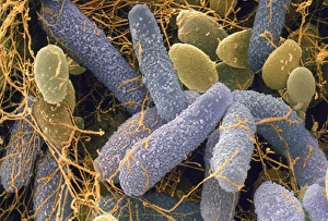
Escherishia Coli Bacteria / E. Coli - affected by antibiotic Scanning Electron Micrograph (SEM): Magnification x25, 000 (if print A4 size)

Background imageBacteria Collection: Anton van Leeuwenhoek, caricature

Anton van Leeuwenhoek, caricature
Anton van Leeuwenhoek. Caricature of the Dutch microbiologist Anton van Leeuwenhoek (1632-1723), looking through a magnifying glass to represent his discoveries in microbiology

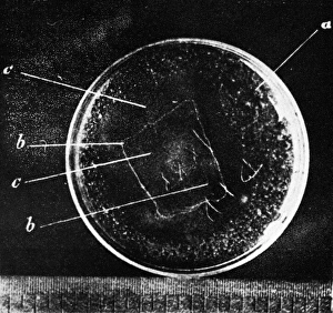
Background imageBacteria Collection: Antibiotic action, historical image

Antibiotic action, historical image
Antibiotic action. Historical image taken by the German pathologist Karl Dohle (1855-1928) in 1889, showing the action of an antibiotic compound on Bacillus anthracis (anthrax)

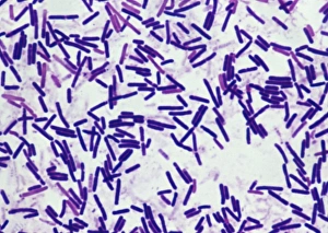
Background imageBacteria Collection: Botulism bacteria

Botulism bacteria. Light micrograph of botulism bacteria (Clostridium botulinum). These bacteria occur naturally in soil. They produce botulinum toxin

Background imageBacteria Collection: Grand Prismatic Spring at Midway Geyser Basin, Yellowstone National Park, Teton County, Wyoming, USA

Grand Prismatic Spring at Midway Geyser Basin, Yellowstone National Park, Teton County, Wyoming, USA

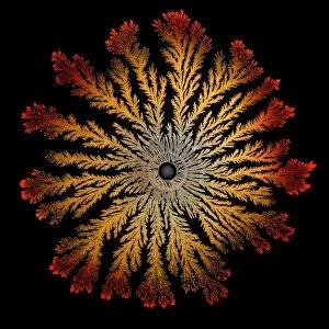
Background imageBacteria Collection: Orbicular Design

Orbicular Design

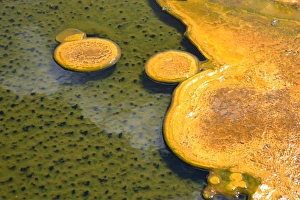
Background imageBacteria Collection: Paint Pots of Yellowstone National Park

Paint Pots of Yellowstone National Park
A close up view of vivid, orange-colored bacteria mats floating in hot springs at Yellowstone National Park. These features are called paint pots in the park

Background imageBacteria Collection: vertical, pool, geyser, heat, yellowstone national park, landscape, scenics, awe

vertical, pool, geyser, heat, yellowstone national park, landscape, scenics, awe
Black Pool in West Thumb Geyser Basin, Yellowstone National Park, Wyoming

Background imageBacteria Collection: Bacillus bacterial genus

Bacillus bacterial genus

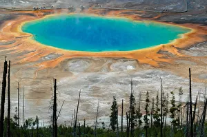
Background imageBacteria Collection: The multi-colored Grand Prismatic spring in Yellowstone National Park as seen from above

The multi-colored Grand Prismatic spring in Yellowstone National Park as seen from above
The Grand Prismatic Spring in Yellowstone National Park is the largest hot spring in the United States, and the third largest in the world

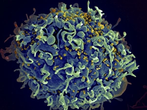
Background imageBacteria Collection: Scanning electron micrograph of HIV particles infecting a human H9 T cell

Scanning electron micrograph of HIV particles infecting a human H9 T cell

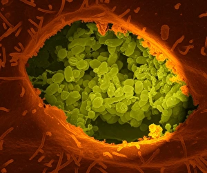
Background imageBacteria Collection: Coxiella burnetii, the bacteria that causes Q Fever

Coxiella burnetii, the bacteria that causes Q Fever. A dry fracture of a Vero cell exposing the contents of a vacuole where Coxiella burnetii are busy growing

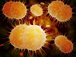
Background imageBacteria Collection: Microscopic view of Neisseria gonorrhoeae

Microscopic view of Neisseria gonorrhoeae. Neisseria gonorrhoeae typically infects the mucous membranes causing infections such as urethritis, cervicitis, salpingitis, pelvic inflammatory disease

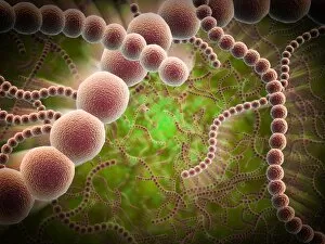
Background imageBacteria Collection: Microscopic view of bacterial pneumonia

Microscopic view of bacterial pneumonia. Bacterial pneumonia is a type of pneumonia caused by bacterial infection. Pneumonia can be generally defined as inflammation of the lung parenchyma

Background imageBacteria Collection: Microscopic view of Streptococcus pneumoniae

Microscopic view of Streptococcus pneumoniae, also known as pneumococcus. Pneumococcus is a Gram-positive coccus shaped pathogenic bacteria which causes many types of pneumococcal infections in

Background imageBacteria Collection: Picture No. 11014633

Picture No. 11014633
Penicillin Colony This is a culture of the original strain of bacteria used by Flemming in the early research into the first antibiotics. Obliquely lit macro image from a time-lapse sequence. Date:
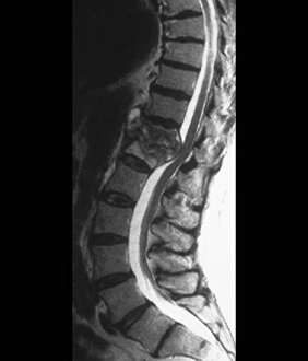
Background imageBacteria Collection: Tuberculosis of the spine, MRI scan

Tuberculosis of the spine, MRI scan
Tuberculosis of the spine. Magnetic resonance imaging (MRI) scan of a sagittal section through the spine of a 74-year-old patient with tuberculosis (TB) of the spine, or Potts disease

Background imageBacteria Collection: MscL ion channel protein structure

MscL ion channel protein structure. Molecular model showing the protein structure of a Mechanosensitive Channel of Large Conductance (MscL) from a Mycobacterium tuberculosis bacterium

Background imageBacteria Collection: Propionibacterium acnes C014 / 0631

Propionibacterium acnes C014 / 0631
Propionibacterium acnes bacteria, coloured scanning electron micrograph (SEM). This bacteria is found deep in the hair follicles and pores of the skin

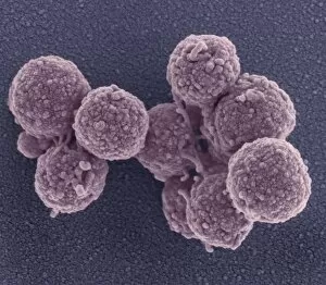
Background imageBacteria Collection: Synthetic Mycoplasma bacteria, SEM C013 / 4777

Synthetic Mycoplasma bacteria, SEM C013 / 4777
Synthetic Mycoplasma bacteria. Coloured scanning electron micrograph (SEM) of Mycoplasma mycoides JCVI-syn1.0 cells. This was the first self-replicating bacterium controlled by a synthetic genome

Background imageBacteria Collection: Coloured SEM of Bacillus sp. bacteria

Coloured SEM of Bacillus sp. bacteria
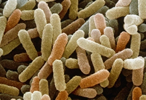
Bacillus sp. Coloured scanning electron micrograph of bacteria cells (Bacillus sp.). These rod- shaped bacteria are found in soil and water and are usually saprotrophic

Background imageBacteria Collection: Acetobacter and Schizosaccharomyces

Acetobacter and Schizosaccharomyces. Coloured scanning electron micrograph of Acetobacter bacteria and Schizosaccharomyces yeast cells in a culture of Kombucha" tea

Background imageBacteria Collection: Anthrax antibiotics research

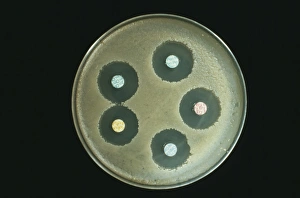
Anthrax antibiotics research. Antibiotic drugs (discs) being tested on anthrax bacteria (Bacillus anthracis) colonies being cultured on an agar growth medium in a petri dish

Background imageBacteria Collection: Rod shaped bacillus bacteria

Rod shaped bacillus bacteria
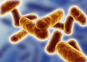
3D-computer artwork of bacillus bacteria. These rod-shaped bacteria (bacilli) are widespread in soil and in the air. Many bacillus bacteria are responsible for food spoilage

Background imageBacteria Collection: Bacteria research

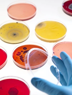
Bacteria research
MODEL RELEASED. Bacteria research. Researcher holding an empty petri dish in front of other dishes containing cultured bacteria (dots)

Background imageBacteria Collection: Chert rock

Chert rock. This carbonaceous chert is from the Barberton greenstone belt in South Africa. The Earths oldest fossil bacteria have been found in this type of rock

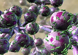
Background imageBacteria Collection: Bacterial lung infection

Bacterial lung infection, computer artwork. Rod-shaped bacteria (bacilli, green) on the surface of the alveoli of the lungs (purple)

Background imageBacteria Collection: Bacillus subtilis bacteria, SEM

Bacillus subtilis bacteria, SEM
Bacillus subtilis. Coloured scanning electron micrograph (SEM) of Bacillus subtilis bacteria. B. subtilis, or hay bacillus, is an aerobic, gram-positive bacillus(rod-shaped bacterium)

Background imageBacteria Collection: Lactobacillus bacteria, SEM

Lactobacillus bacteria, SEM
Lactobacillus sp. bacteria, coloured scanning electron micrograph (SEM). Lactobacillus bacteria are probiotic bacteria, which are useful for human and animal health as they produce bacteriocins

Background imageBacteria Collection: Clostridium perfringens bacterium with spore

Clostridium perfringens bacterium with spore
False-colour transmission electron micrograph (TEM) of a Clostridium perfringens bacterium with endospore. This species of Gram-positive

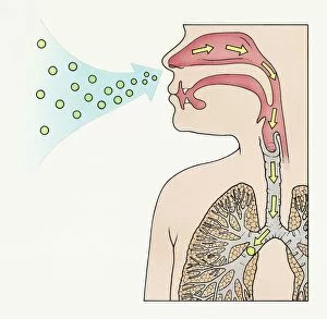
Background imageBacteria Collection: Bacteria on lung hairs

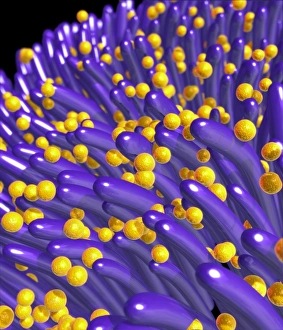
Bacteria on lung hairs
Bacteria in a lung. Computer artwork of bacteria (yellow) on cilia (hair-like structures) in a lung. Bacterial infection of the lungs can cause potentially fatal pneumonia

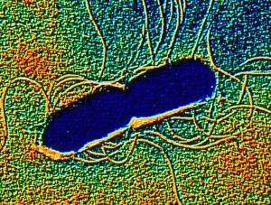
Background imageBacteria Collection: Proteus mirabilis bacterium

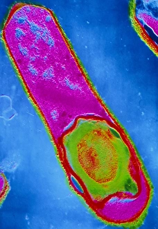
Proteus mirabilis bacterium
False-colour transmission electron micrograph of the bacterium Proteus mirabilis. It is an enterobacterium which is present normally in the human intestine

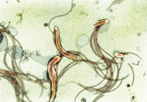
Background imageBacteria Collection: Spirochete bacteria, TEM

Spirochete bacteria, TEM
Spirochete bacteria, coloured transmission electron micrograph (TEM). Spirochetes are bacteria with helically coiled cells

Background imageBacteria Collection: Coloured SEM of Clostridium botulinum

Coloured SEM of Clostridium botulinum
Food poisoning bacterium. Coloured scanning electron micrograph of the Gram-positive anaerobic bacteria Clostridium botulinum, responsible for serious food poisoning in humans. C

Background imageBacteria Collection: Rod-shaped bacteria

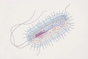
Rod-shaped bacteria. Computer artwork of rod- shaped bacteria (bacilli). Typical bacilli are E. coli and Salmonella bacteria, though there are many others as well

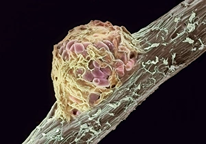
Background imageBacteria Collection: Root nodule

Root nodule. Coloured scanning electron micrograph (SEM) of a root nodule on a pea plant (Pisum sativum) caused by the nitrogen-fixing soil bacteria Rhizobium leguminosarum

Background imageBacteria Collection: A colony of bacteria seen under a microscope

A colony of bacteria seen under a microscope

Background imageBacteria Collection: bacteria

bacteria
microscopic image

Background imageBacteria Collection: Fumigating a Room

Fumigating a Room
This young woman covers her mouth to protect herself from breathing in harmful bacteria as she seals a doorway and keyhole of a room in her house for fumigation from disease. Date: early 1930s

Background imageBacteria Collection: Advertisement for Salutaris bottled table water

Advertisement for Salutaris bottled table water, pure, distilled and aerated -- no foreign bodies permitted. Date: early 20th century

Background imageBacteria Collection: Pattern in Canary Spring, Yellowstone National Park, Montana / Wyoming

Pattern in Canary Spring, Yellowstone National Park, Montana / Wyoming
Pattern in Canary Spring, Yellowstone National Park (Montana, Wyoming)

Background imageBacteria Collection: A diagram showing aspergilloma affecting human lung

A diagram showing aspergilloma affecting human lung

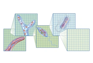
Background imageBacteria Collection: Cross section biomedical illustration on grid of Worm, Fungi, Protozoa

Cross section biomedical illustration on grid of Worm, Fungi, Protozoa, and Bacteria infection and infestation

Background imageBacteria Collection: Picture No. 11675606

Picture No. 11675606
Patterns of Paenibacillus bacteria on petri dish - T type (tip-splitting morphotype). Date:

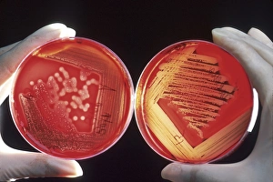
Background imageBacteria Collection: Red blood cells on an agar plate are used to diagnose infection

Red blood cells on an agar plate are used to diagnose infection. The plate on the left shows a positive staphyloccus infection

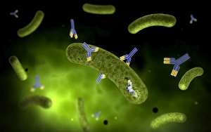
Background imageBacteria Collection: Conceptual image of antibody attaching and killing bacteria

Conceptual image of antibody attaching and killing bacteria


All Professionally Made to Order for Quick Shipping

"Bacteria: The Microscopic Marvels of Yellowstone National Park" In the heart of Wyoming, USA lies the breathtaking Yellowstone National Park, a haven for nature enthusiasts and scientists alike. Amidst its natural wonders, one can find an extraordinary display of microbial life at the Grand Prismatic Spring in the Midway Geyser Basin. Picture No. 11675590 captures this mesmerizing sight, showcasing vibrant hues that seem to dance upon the water's surface. However, what may appear as an artistic masterpiece is actually a thriving community of bacteria. Zooming in closer with SEM C018 / 8596 reveals a neutrophil engulfing MRSA - Methicillin-resistant Staphylococcus aureus. This image serves as a reminder of how these tiny organisms can pose significant threats to human health. Another SEM image showcases E. Coli bacteria, reminding us that not all it can harmful; some play crucial roles in our digestive system and even aid scientific research. Moving back to Yellowstone's enchantment, we encounter Salmonella bacteria through another captivating SEM photograph. These microscopic creatures serve as a stark reminder that even within such serene landscapes, dangers lurk unseen. Coloured TEM imagery introduces us to Yersinia pestis bacteria – responsible for causing devastating outbreaks like the infamous Black Death throughout history. Such visuals emphasize their intricate structures and highlight why understanding them is vital for public health efforts. Venturing beyond real-life images into computer artwork brings forth a beta DNA segment surrounded by spheres—a representation of genetic material essential for bacterial survival and reproduction. Artistic depictions also shed light on various cell types found within these microorganisms' complex world—each playing unique roles necessary for their survival and proliferation. Historical diagrams reveal cultures of Anthrax—an infectious disease with severe consequences if left unchecked—underscoring humanity's ongoing battle against bacterial infections throughout time. Tuberculosis bacteria make their appearance too—a haunting reminder that despite medical advancements, infectious diseases continue to challenge us.