
Atheroma Collection
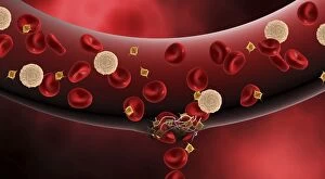
Atheroma, also known as plaque buildup in the arteries, is a common condition that can lead to serious health complications


All Professionally Made to Order for Quick Shipping
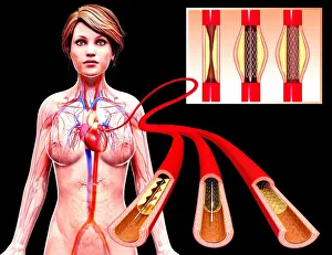
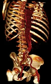
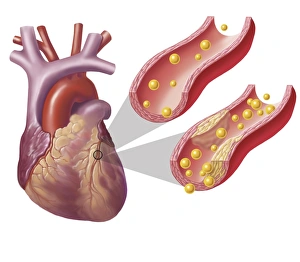
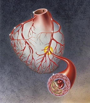
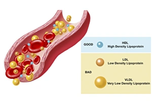
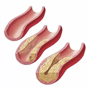
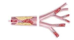
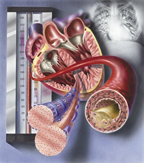
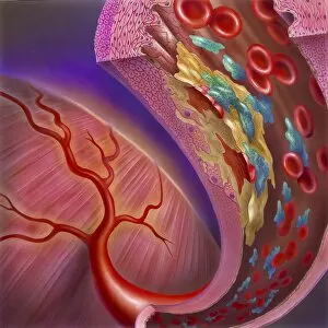
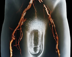
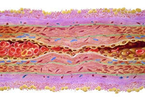
Atheroma, also known as plaque buildup in the arteries, is a common condition that can lead to serious health complications. Angioplasty, a procedure used to widen narrowed or blocked arteries, has become an effective treatment option for patients with atheroma. Computer artwork depicting this medical intervention showcases the intricate process of restoring blood flow and preventing further damage. In contrast, computer artwork illustrating a blocked artery highlights the severity of this condition, and are obstruct blood flow to vital organs such as the heart and brain, increasing the risk of heart attacks and strokes. The visual representation serves as a reminder of the importance of early detection and timely intervention. Another potential consequence is an aortic dissection – when there is tearing in the inner layer of the body's largest artery (the aorta). 3D CT scans provide detailed images that aid in diagnosing this life-threatening condition accurately. By identifying an aortic dissection promptly through these advanced imaging techniques, doctors can initiate appropriate treatment strategies swiftly. The series of pictures numbered 11675562 to 11675554 depict various aspects related to understanding and managing atheroma effectively. These visuals offer valuable insights into its impact on our cardiovascular system while emphasizing modern medical advancements like angioplasty and diagnostic tools like CT scans. As we continue to explore innovative approaches towards combating diseases like atheroma, it becomes crucial for individuals to prioritize their cardiovascular health through regular check-ups and adopting healthy lifestyle choices.