
Articulation Collection
"Exploring the Intricacies of Articulation: From Ankle Ligaments to Turkish Belly Dance" Articulation, a fascinating aspect of human anatomy and movement


All Professionally Made to Order for Quick Shipping
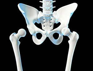
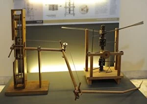
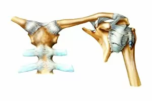
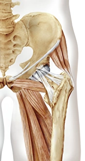
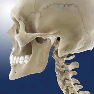
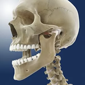
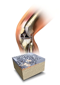
"Exploring the Intricacies of Articulation: From Ankle Ligaments to Turkish Belly Dance" Articulation, a fascinating aspect of human anatomy and movement, encompasses various joints and ligaments throughout our body. Delving into this intricate subject reveals a world of complexity and beauty. Starting with the ankles, we encounter the outer ankle ligaments (artwork C013 / 4452) and inner ankle ligaments (artwork C013 / 4451). These delicate structures play a crucial role in providing stability while allowing for fluid motion. Moving on to Le Vocabulaire Illustre: Articulation; Knochenfugung (engraving), we witness detailed depictions that showcase the artistry behind articulations. Leonardoesque models further captivate us with their adjustable-tilt wing and wing-beating mechanisms, reminding us of how nature inspires innovation (Leonardesque models. Adjustable-tilt wing and Wing-beating d). Stepping away from anatomical illustrations, we find ourselves immersed in the enchanting realm of Turkish belly dance. This mesmerizing dance form showcases remarkable articulation skills as performers effortlessly move their bodies with grace and precision. Returning to our exploration of joints, hip joint bones take center stage alongside artwork C014 / 2032's depiction of their anatomy. The intricacy involved in these connections is further highlighted by an engraving from 1866 titled "Articulations anatomy. " Shifting focus towards hip bones (artwork C016 / 7015), we gain insight into how these sturdy structures contribute to our overall mobility. Additionally, vertebral joints within the neck region (artwork C016 / 6551) intrigue us with their flexibility while maintaining stability. Lastly, we encounter two vital junctions.