
Anther Collection (page 7)
"Exploring the Intricate World of Anthers: A Captivating Microscopic Journey" Delve into the mesmerizing realm of anthers


All Professionally Made to Order for Quick Shipping
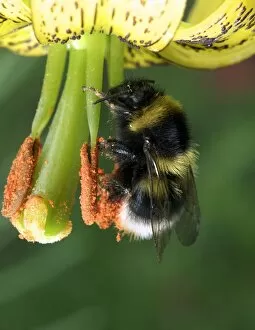
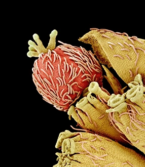
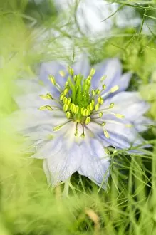
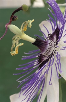
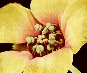
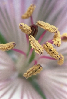
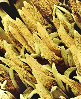
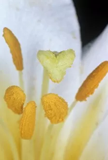
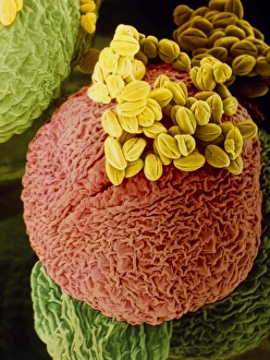
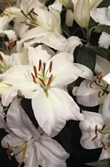
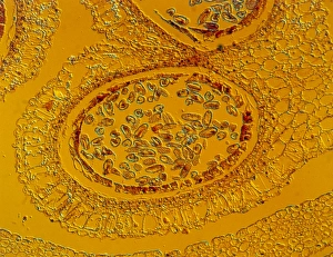
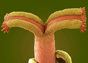
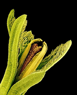
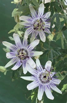
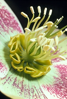
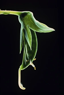
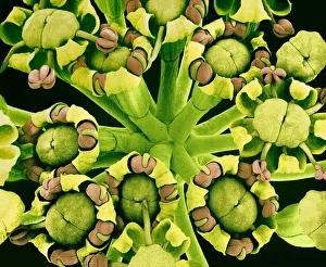
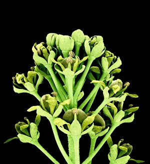
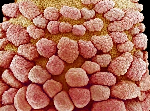
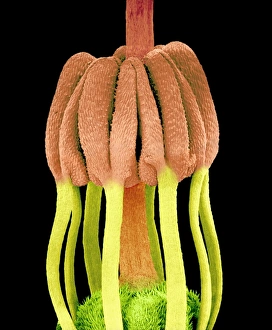
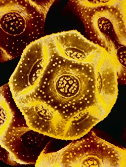
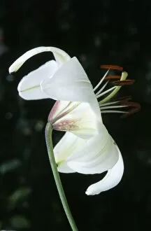
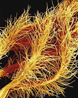
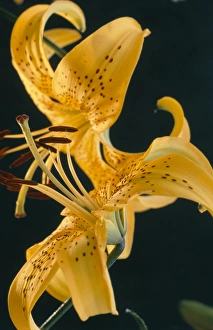
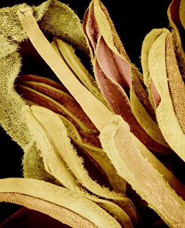
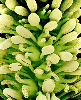
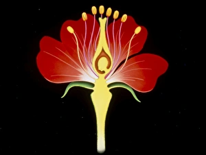
"Exploring the Intricate World of Anthers: A Captivating Microscopic Journey" Delve into the mesmerizing realm of anthers, where nature's intricate beauty unfolds at a microscopic level. From the vibrant Flame lily (Gloriosa sp. ) to the delicate Geranium anther captured in stunning detail through scanning electron microscopy (SEM), this captivating journey takes us deep within the heart of flowers. Witness the Tea flower stamens as they reveal their enchanting structures under SEM, showcasing their unique patterns and textures. The Geranium pollen, magnified to perfection, showcases its fine intricacies that aid in pollination and reproduction. Marvel at the Forget-me-not flower's SEM image, unveiling its ethereal petals and stamen arrangement with unparalleled clarity. The Hong Kong orchid tree (Bauhinia blakeana) flowers from December bloom with grace and elegance, inviting us to appreciate their exquisite form. The Buttercup flower's SEM image reveals a world of miniature wonders hidden within its golden petals. Delicate Columbine flower stamens come alive under high-resolution SEM imaging, displaying their remarkable shapes and colors. A false-color SEM image captures the essence of a chickweed flower like never before—a visual feast for our senses. The majestic Amaryllis (Hippeastrum sp. ) stands tall with its resplendent blooms that captivate all who behold them. Finally, we encounter Thale cress flower through a micrograph—an intimate glimpse into nature's meticulous design process on a cellular scale. These diverse anthers remind us of Mother Nature's boundless creativity and her attention to even the tiniest details. Embark on this extraordinary expedition into anther exploration; let these images transport you into a world unseen by naked eyes but brimming with awe-inspiring beauty waiting to be discovered.