Fraud Blocker Skip to main content

Anterior View Collection

"Exploring the Intricacies of the Anterior View

Background imageAnterior View Collection: Anterior view total shoulder joint repair

Anterior view total shoulder joint repair
Horizontal, Detail, Anatomy, Diagram, Repair, Illustration, Bone, Shoulder, Biology, Joint, Cut Out, White Background, Artwork, Color Image, Front View, Close Up, Healthcare And Medicine

Background imageAnterior View Collection: White matter fibres, DTI scan

White matter fibres, DTI scan
White matter fibres. Coloured 3D diffusion tensor imaging (DTI) scan of the brain, showing the bundles of white matter nerve fibres

Background imageAnterior View Collection: Anterior view of pelvis with hip bones showing arthritis and osteophytes on femoral heads

Anterior view of pelvis with hip bones showing arthritis and osteophytes on femoral heads
Horizontal, Anatomy, Diagram, Illustration, Bone, Disease, Medical, Hip, Biology, Pelvis, Femur, Cut Out, Arthritis, White Background, Artwork, Color Image, Front View, Healthcare And Medicine

Background imageAnterior View Collection: Normal front view of the adult skull

Normal front view of the adult skull
Anatomy, Diagram, Illustration, Bone, Teeth, Skull, Medical, Biology, Cut Out, White Background, Vertical, Artwork, Color Image, Front View, Human Skull, Healthcare And Medicine, The Human Body

Background imageAnterior View Collection: Normal anterior view of pelvis with hip bones

Normal anterior view of pelvis with hip bones
Horizontal, Anatomy, Diagram, Illustration, Bone, Medical, Hip, Biology, Pelvis, Femur, Cut Out, White Background, Artwork, Color Image, Front View, Healthcare And Medicine, The Human Body

Background imageAnterior View Collection: Repetitive activities like aerobics that involve lifting up the knee can cause trauma to

Repetitive activities like aerobics that involve lifting up the knee can cause trauma to
Detail, Anatomy, Diagram, Muscle, Pain, Illustration, Bone, Trauma, Injury, Medical, Biology, Pelvis, Femur, See Through, Cut Out, White Background, Artwork, Color Image, Front View, Close Up

Background imageAnterior View Collection: Anterior view showing normal versus enlarged prostate gland

Anterior view showing normal versus enlarged prostate gland
Horizontal, Detail, Anatomy, Diagram, Illustration, Contrast, Medical, Biology, Abnormal, Anterior, Cut Out, White Background, Artwork, Cross Section, Color Image, Front View, Close Up

Background imageAnterior View Collection: Front view of a normal heart and its cornonary arteries and veins

Front view of a normal heart and its cornonary arteries and veins
Detail, Anatomy, Diagram, Heart, Illustration, Medical, Vein, Biology, Cardiac, Veins, Cardiovascular, Cut Out, White Background, Vertical, Artwork, Color Image, Front View, Close Up

Background imageAnterior View Collection: Normal anterior view of a pelvis

Normal anterior view of a pelvis
Horizontal, Anatomy, Diagram, Illustration, Bone, Medical, Hip, Biology, Pelvis, Cut Out, White Background, Artwork, Color Image, Front View, Healthcare And Medicine, The Human Body, No People

Background imageAnterior View Collection: Anterior view of a normal uterus with ovaries, fallopian tubes, and broad ligament

Anterior view of a normal uterus with ovaries, fallopian tubes, and broad ligament
Horizontal, Detail, Anatomy, Diagram, Illustration, Medical, Uterus, Biology, Reproduction, Normal, Cut Out, White Background, Artwork, Female Likeness, Color Image, Front View, Close Up

Background imageAnterior View Collection: Anatomical Print of the Human Skeleton and Muscles, 1935 (screen print)

Anatomical Print of the Human Skeleton and Muscles, 1935 (screen print)
3035954 Anatomical Print of the Human Skeleton and Muscles, 1935 (screen print) by American School, (20th century); (add.info)

Background imageAnterior View Collection: Normal coronal section of the skull and brain showing the coronal sinuses

Normal coronal section of the skull and brain showing the coronal sinuses
Brain, Anatomy, Diagram, Illustration, Bone, Skull, Medical, Eyeball, Biology, See Through, Cut Out, White Background, Eye, Vertical, Artwork, Color Image, Front View, Human Skull

Background imageAnterior View Collection: Front view of a body showing a total hip replacement

Front view of a body showing a total hip replacement
Detail, Anatomy, Diagram, Illustration, Bone, Disease, Medical, Hip, Biology, Pelvis, Femur, Acetabulum, Surgery, See Through, Cut Out, White Background, Vertical, Artwork, Color Image, Front View

Background imageAnterior View Collection: Anterior view female anatomy showing breast tissue with a tumor

Anterior view female anatomy showing breast tissue with a tumor
Anatomy, Diagram, Illustration, Medical, Biology, Surgery, Cancer, See Through, Cut Out, White Background, Vertical, Artwork, Female Likeness, Breast, Color Image, Front View

Background imageAnterior View Collection: Anterior view of legs and the veinous system

Anterior view of legs and the veinous system
Detail, Anatomy, Diagram, Illustration, Standing, Leg, Barefoot, Medical, Vein, Biology, See Through, Cut Out, White Background, Vertical, Artwork, Low Section, Color Image, Front View, Close Up

Background imageAnterior View Collection: Armpit muscles, 1831 artwork

Armpit muscles, 1831 artwork
Armpit muscles. Muscle connections in the second layer of the armpit. This anatomical artwork is plate 110 from volume 2 (1831) of Traite complet de l anatomie de l homme (1831-1854)

Background imageAnterior View Collection: Odontaspis robusta, sand tiger shark teeth

Odontaspis robusta, sand tiger shark teeth
Odontaspis robusta from the Early Eocene rocks of Abbey Wood, Kent. Anterior teeth (tall, slender) and lateral teeth (triangular) are shown

Background imageAnterior View Collection: Fascia of the lower leg, 1831 artwork

Fascia of the lower leg, 1831 artwork
Fascia of the lower leg, anterior (left) and posterior (right) views. Fascia are fibrous connective tissues that surround muscles and other tissues

Background imageAnterior View Collection: Face and neck muscles, 1831 artwork

Face and neck muscles, 1831 artwork
Face and neck muscle. Anterior view of the deep layers of muscles of the face and neck, including the eyes and mouth. Insets show details of the lips, chin and eyes

Background imageAnterior View Collection: Neck muscle anatomy, 1831 artwork

Neck muscle anatomy, 1831 artwork
Neck muscle anatomy. Various views of the anatomy of the muscles of the neck. The main artwork shows the neck seen from the front

Background imageAnterior View Collection: Trunk muscle anatomy, 1831 artwork

Trunk muscle anatomy, 1831 artwork
Trunk muscle anatomy. Coronal section of the trunk (anterior view) showing the anatomy of its muscles and bones. This anatomical artwork is plate 75 from volume 2 (1831)

Background imageAnterior View Collection: Trunk musculature, 1831 artwork

Trunk musculature, 1831 artwork
Trunk musculature, anterior view. This anatomical artwork is plate 62 from volume 2 (1831) of Traite complet de l anatomie de l homme (1831-1854)

Background imageAnterior View Collection: Whole body musculature, 1831 artwork

Whole body musculature, 1831 artwork
Whole body musculature, anterior view. This anatomical artwork is plate 60 from volume 2 (1831) of Traite complet de l anatomie de l homme (1831-1854)

Background imageAnterior View Collection: Thigh muscle anatomy, 1831 artwork

Thigh muscle anatomy, 1831 artwork
Thigh muscle anatomy. Anterior view of some of the layers of the muscles of the thigh in relation to the femur (thigh bone) and the pelvis bones

Background imageAnterior View Collection: Groin muscles, 1831 artwork

Groin muscles, 1831 artwork
Groin muscles. Muscles of the anterior wall of the lower (groin) region of the trunk, with a study of the inguinal canal. This anatomical artwork is plate 70 from volume 2 (1831)

Background imageAnterior View Collection: Homo sapiens, human and Gorilla gorilla, western gorilla

Homo sapiens, human and Gorilla gorilla, western gorilla
Drawing of a human skeleton and a gorilla skeleton. Published in Transactions of the Zoological Society of London Vol. 5, Part 1 by Richard Owen, 1866

Background imageAnterior View Collection: Mesosaurus tenuidens fossil

Mesosaurus tenuidens fossil
Anterior portion of skeleton of the extinct reptile, Mesosaurus tenuidens gervais from the Karoo Formation, Griqualand West, South Africa. Cast of the type specimen

Background imageAnterior View Collection: Deglutition in Chauliodus

Deglutition in Chauliodus
Illustration of main movements of the anterior part of the body and the head of Chauliodus when it catches and swallows large prey

Background imageAnterior View Collection: Ovis vignei vignei, Ladakh urial

Ovis vignei vignei, Ladakh urial
Frontal view of skull with horns of Ovis vignei vignei, a urial wild sheep, from the Ladakh region of India

Background imageAnterior View Collection: Osteological Gallery, Natural History Museum, London, July 1

Osteological Gallery, Natural History Museum, London, July 1

Background imageAnterior View Collection: Respiratory system

Respiratory system, anterior (front) view. Artwork taken from The Viscera of the Human Body (1840), a textbook edited by Jones Quain and Erasmus Wilson

Background imageAnterior View Collection: Knee in anterior view, artwork

Knee in anterior view, artwork
Artwork of the anatomy of the knee joint (anterior view) showing the femur (thigh bone, at top) articulating with the tibia (shin bone, at bottom), cartilage (light blue)

Background imageAnterior View Collection: Knee joint anatomy, artwork

Knee joint anatomy, artwork
Artwork of the anatomy of the knee joint (anterior view) showing the femur (thigh bone, at top) articulating with the tibia (shin bone, at bottom), articular cartilage

Background imageAnterior View Collection: Cruciate ligament injury, artwork

Cruciate ligament injury, artwork
Artwork (anterior view) of the anatomy of the knee joint showing the inner cruciate ligaments (purple) arranged like a cross which serve to stabilize the knee joint while allowing motion

Background imageAnterior View Collection: Sports injuries to the groin, artwork

Sports injuries to the groin, artwork
Artwork (anterior view) of the anatomy of the hip and thigh showing muscles and their associated bones which most commonly suffer sports injuries to the groin

Background imageAnterior View Collection: Heart nerves, 1844 artwork

Heart nerves, 1844 artwork
Heart nerves. This anatomical artwork is figure 3, plate 98 from volume 3 (1844) of Traite complet de l anatomie de l homme (1831-1854)

Background imageAnterior View Collection: Eye anatomy, 1844 artwork

Eye anatomy, 1844 artwork
Eye anatomy, front view. This anatomical artwork is figure 2, plate 75 from volume 3 (1844) of Traite complet de l anatomie de l homme (1831-1854)

Background imageAnterior View Collection: Spinal cord anatomy, 1844 artwork

Spinal cord anatomy, 1844 artwork
Spinal cord anatomy. These anatomical artworks form plate 11 from volume 3 (1844) of Traite complet de l anatomie de l homme (1831-1854)


All Professionally Made to Order for Quick Shipping

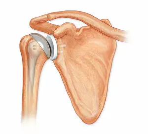
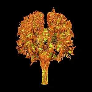
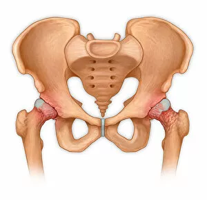
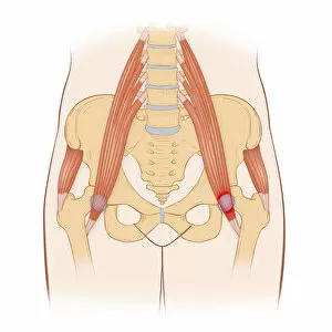
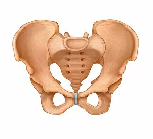
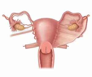
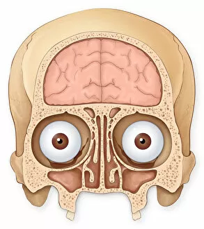
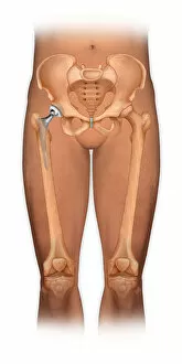
"Exploring the Intricacies of the Anterior View: From Shoulder Joint Repair to Brain Anatomy" An anterior view total shoulder joint repair reveals the intricate process of restoring mobility and function. Delving into white matter fibres through a DTI scan unravels the complex network that facilitates communication within our brain. A captivating 1831 artwork showcases the detailed depiction of face and neck muscles, highlighting their importance in expression and movement. Witnessing a normal anterior view of pelvis with hip bones provides insight into its structure and alignment crucial for proper locomotion. Repetitive activities like aerobics involving knee lifting can lead to trauma, emphasizing the need for caution during exercise routines. Comparing an anterior view shows both normal and enlarged prostate gland, shedding light on potential health concerns affecting men's well-being. Arthritis and osteophytes on femoral heads become evident when observing an anterior view of pelvis with hip bones, underlining degenerative conditions that impact mobility. Gaining knowledge about a normal front view of an adult skull allows us to appreciate its intricacy as it protects our most vital organ – the brain. Examining a normal anterior view of a pelvis helps understand its role in supporting reproductive organs while maintaining stability during movement. An intriguing glimpse at an anatomical print from 1935 unveils the human skeleton's complexity alongside muscular structures, showcasing how they work together harmoniously. A normal coronal section displaying skull and brain anatomy highlights essential features such as coronal sinuses critical for drainage functions.