
Anterior Cruciate Ligament Collection
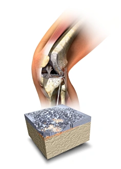
The anterior cruciate ligament (ACL) is a vital component of the human knee joint


All Professionally Made to Order for Quick Shipping
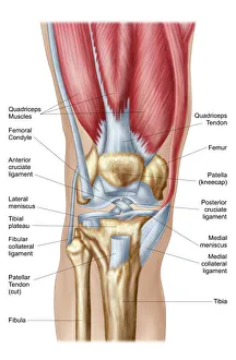
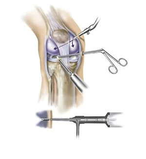
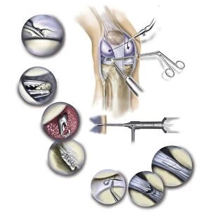
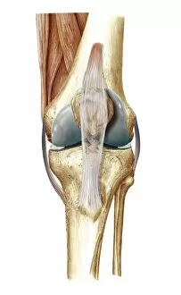
The anterior cruciate ligament (ACL) is a vital component of the human knee joint. It plays a crucial role in stabilizing the knee and preventing excessive forward movement of the tibia bone. This intricate anatomy can be seen in a detailed cutaway illustration of the human knee, showcasing its complexity. During arthroscopic surgical procedures, specialized instruments are inserted into the knee to repair or reconstruct the ACL. The insertion points for these instruments can be observed in a close-up view of the knee, highlighting how surgeons navigate this delicate area. As part of a typical synovial joint, which allows for smooth movement between bones, the ACL works alongside other ligaments and structures to maintain stability and flexibility. Artwork depicting knee bones and ligaments further emphasizes their interconnection. Medical tests such as the Lachman test help diagnose ACL injuries by assessing abnormal movements within the joint. In cases where an ACL tear is confirmed through imaging techniques like X-ray or CT scan, prompt treatment becomes essential to restore function and prevent long-term complications. Understanding the intricacies of our knees' anatomy helps us appreciate how important it is to protect our anterior cruciate ligament from injury or damage that could impact our mobility and overall quality of life.