Fraud Blocker Skip to main content

Anatomical Chart Collection

EDITORS COMMENTS"Exploring the Intricacies of Human and Animal Anatomy

Background imageAnatomical Chart Collection: Anatomical chart, engraving by Ercole Lelli (1702-1766)

Anatomical chart, engraving by Ercole Lelli (1702-1766)
DGA1257595 Anatomical chart, engraving by Ercole Lelli (1702-1766) by Lelli, Ercole (1702-66); (add.info.: Anatomical chart)

Background imageAnatomical Chart Collection: Shi si jing fa hui

Shi si jing fa hui
2626560 Shi si jing fa hui; (add.info.: Illustration from the Japanese & Chinese edition (1716) of Shi si jing fa hui

Background imageAnatomical Chart Collection: Shi si jing fa hui

Shi si jing fa hui
2626541 Shi si jing fa hui; (add.info.: Illustration from the Japanese & Chinese edition (1716) of Shi si jing fa hui

Background imageAnatomical Chart Collection: Shi si jing fa hui

Shi si jing fa hui
2626557 Shi si jing fa hui; (add.info.: Illustration from the Japanese & Chinese edition (1716) of Shi si jing fa hui

Background imageAnatomical Chart Collection: Shi si jing fa hui

Shi si jing fa hui
2626545 Shi si jing fa hui; (add.info.: Illustration from the Japanese & Chinese edition (1716) of Shi si jing fa hui

Background imageAnatomical Chart Collection: Traite de medecine de Mansour : Muscle et systeme nerveux humain

Traite de medecine de Mansour : Muscle et systeme nerveux humain
AIS5373969 Traite de medecine de Mansour : Muscle et systeme nerveux humain. Manuscrit perse

Background imageAnatomical Chart Collection: Normal disposition of the viscera in the human body

Normal disposition of the viscera in the human body
7295202 Normal disposition of the viscera in the human body. "Life and health" by Dr Jules Rengade. Paris 1881; Private Collection; Photo ©Jaime Abecasis

Background imageAnatomical Chart Collection: Shi si jing fa hui

Shi si jing fa hui
2626543 Shi si jing fa hui; (add.info.: Illustration from the Japanese & Chinese edition (1716) of Shi si jing fa hui

Background imageAnatomical Chart Collection: Shi si jing fa hui

Shi si jing fa hui
2626558 Shi si jing fa hui; (add.info.: Illustration from the Japanese & Chinese edition (1716) of Shi si jing fa hui

Background imageAnatomical Chart Collection: Shi si jing fa hui

Shi si jing fa hui
2626540 Shi si jing fa hui; (add.info.: Illustration from the Japanese & Chinese edition (1716) of Shi si jing fa hui

Background imageAnatomical Chart Collection: Digestive tract. The caecum or cecum and the appendix ileo cecal

Digestive tract. The caecum or cecum and the appendix ileo cecal
7295213 Digestive tract. The caecum or cecum and the appendix ileo cecal. Book by Dr Jules Rengade "Life and Health", Paris 1881; Private Collection; Photo ©Jaime Abecasis

Background imageAnatomical Chart Collection: Hematosis and blood circulation

Hematosis and blood circulation
7295210 Hematosis and blood circulation. "Life and health " by Dr Jules Rengade, Paris 1881; Private Collection; Photo ©Jaime Abecasis

Background imageAnatomical Chart Collection: Shi si jing fa hui

Shi si jing fa hui
2626565 Shi si jing fa hui; (add.info.: Illustration from the Japanese & Chinese edition (1716) of Shi si jing fa hui

Background imageAnatomical Chart Collection: Shi si jing fa hui

Shi si jing fa hui
2626563 Shi si jing fa hui; (add.info.: Illustration from the Japanese & Chinese edition (1716) of Shi si jing fa hui

Background imageAnatomical Chart Collection: Shi si jing fa hui

Shi si jing fa hui
2626544 Shi si jing fa hui; (add.info.: Illustration from the Japanese & Chinese edition (1716) of Shi si jing fa hui

Background imageAnatomical Chart Collection: Shi si jing fa hui

Shi si jing fa hui
2626539 Shi si jing fa hui; (add.info.: Illustration from the Japanese & Chinese edition (1716) of Shi si jing fa hui

Background imageAnatomical Chart Collection: Expression of horror and anguish, 19th century (engraving)

Expression of horror and anguish, 19th century (engraving)
FGL5462706 Expression of horror and anguish, 19th century (engraving); (add.info.: Expression of horror and anguish)

Background imageAnatomical Chart Collection: Animal anatomy: Oyster, lower valve, 1900 (engraving)

Animal anatomy: Oyster, lower valve, 1900 (engraving)
6947883 Animal anatomy: Oyster, lower valve, 1900 (engraving); Private Collection; (add.info.: Animal anatomy: Oyster, lower valve, 1900); Photo ©Jaime Abecasis

Background imageAnatomical Chart Collection: Vertical section of the orbit and the eye ball

Vertical section of the orbit and the eye ball
7295207 Vertical section of the orbit and the eye ball. "Life and Health" by Dr Jules Rengade, Paris, 1881; Private Collection; Photo ©Jaime Abecasis

Background imageAnatomical Chart Collection: Nasal and olfactive nerve. 'Life and Health' by Dr Jules Rengade, Paris, 1881

Nasal and olfactive nerve. "Life and Health" by Dr Jules Rengade, Paris, 1881
7295193 Nasal and olfactive nerve. "Life and Health" by Dr Jules Rengade, Paris, 1881; Private Collection; Photo ©Jaime Abecasis

Background imageAnatomical Chart Collection: Bottom (Fundus) of a normal eye. Right eye, 1902 (published 1930) (print)

Bottom (Fundus) of a normal eye. Right eye, 1902 (published 1930) (print)
7295192 Bottom (Fundus) of a normal eye. Right eye, 1902 (published 1930) (print); Private Collection; Photo ©Jaime Abecasis

Background imageAnatomical Chart Collection: Muscles of the eyelid and the eye ball

Muscles of the eyelid and the eye ball
7295212 Muscles of the eyelid and the eye ball. "Life and health" by Dr Jules Rengade, Paris, 1881; Private Collection; Photo ©Jaime Abecasis

Background imageAnatomical Chart Collection: Venous blood in arterial blood transformation in the lungs cell

Venous blood in arterial blood transformation in the lungs cell
7295211 Venous blood in arterial blood transformation in the lungs cell. Above: end of a bronchial branching

Background imageAnatomical Chart Collection: Shi si jing fa hui

Shi si jing fa hui
2626564 Shi si jing fa hui; (add.info.: Illustration from the Japanese & Chinese edition (1716) of Shi si jing fa hui

Background imageAnatomical Chart Collection: Shi si jing fa hui

Shi si jing fa hui
2626562 Shi si jing fa hui; (add.info.: Illustration from the Japanese & Chinese edition (1716) of Shi si jing fa hui

Background imageAnatomical Chart Collection: Shi si jing fa hui

Shi si jing fa hui
2626561 Shi si jing fa hui; (add.info.: Illustration from the Japanese & Chinese edition (1716) of Shi si jing fa hui

Background imageAnatomical Chart Collection: The Shepherds Calendar, 1480-90 (gold ink & ink on parchment)

The Shepherds Calendar, 1480-90 (gold ink & ink on parchment)
8682933 The Shepherds Calendar, 1480-90 (gold ink & ink on parchment) by French School, (15th century); Fitzwilliam Museum, University of Cambridge

Background imageAnatomical Chart Collection: Human skeleton, front view

Human skeleton, front view
7273837 Human skeleton, front view. by Dupret, S. (19th century); Private Collection; (add.info.: Human skeleton, front view. Set of bones. Anatomical plate drawn by S)

Background imageAnatomical Chart Collection: Illustration from Fasciculus medicinae Venice, 15th century (woodcut)

Illustration from Fasciculus medicinae Venice, 15th century (woodcut)
884991 Illustration from Fasciculus medicinae Venice, 15th century (woodcut) by Italian School, (15th century); Biblioteca Civica, Padua

Background imageAnatomical Chart Collection: Illustration from Fasciculus medicinae Venice, 15th century (woodcut)

Illustration from Fasciculus medicinae Venice, 15th century (woodcut)
884990 Illustration from Fasciculus medicinae Venice, 15th century (woodcut) by Italian School, (15th century); Biblioteca Civica, Padua

Background imageAnatomical Chart Collection: The intestines of a horse. 1792 (engraving)

The intestines of a horse. 1792 (engraving)
7235904 The intestines of a horse. 1792 (engraving) by Dodd, Daniel (fl.1760-90) (after); (add.info.: The intestines of a horse)

Background imageAnatomical Chart Collection: Anatomy of a horse. 1792 (engraving)

Anatomy of a horse. 1792 (engraving)
7235906 Anatomy of a horse. 1792 (engraving) by Dodd, Daniel (fl.1760-90) (after); (add.info.: Anatomy of a horse. Chest cavity exposed 1, heart or vena cava 2, left ventricle 3, and lungs 4)

Background imageAnatomical Chart Collection: Skeleton or bones of a horse. 1792 (engraving)

Skeleton or bones of a horse. 1792 (engraving)
7235907 Skeleton or bones of a horse. 1792 (engraving) by Dodd, Daniel (fl.1760-90) (after); (add.info.: Skeleton or bones of a horse. Handcoloured copperplate engraving by J)

Background imageAnatomical Chart Collection: Anatomy of a horse. 1792 (engraving)

Anatomy of a horse. 1792 (engraving)
7235905 Anatomy of a horse. 1792 (engraving) by Dodd, Daniel (fl.1760-90) (after); (add.info.: Anatomy of a horse. The head, brains, stomach, pancreas, and generative parts)

Background imageAnatomical Chart Collection: Skull, feet and tail of the Eurasian beaver, Castor fiber. 1807 (aquatint)

Skull, feet and tail of the Eurasian beaver, Castor fiber. 1807 (aquatint)
7237456 Skull, feet and tail of the Eurasian beaver, Castor fiber

Background imageAnatomical Chart Collection: Anatomy of bivalve molluscs. 1841 (lithograph)

Anatomy of bivalve molluscs. 1841 (lithograph)
7235256 Anatomy of bivalve molluscs. 1841 (lithograph) by Kull, Conrad (1816-1897); Private Collection; (add.info.: Anatomy of bivalve molluscs. Schultermuscheln)

Background imageAnatomical Chart Collection: Anatomy of jellyfish species. 1841 (lithograph)

Anatomy of jellyfish species. 1841 (lithograph)
7235255 Anatomy of jellyfish species. 1841 (lithograph) by Kull, Conrad (1816-1897); Private Collection; (add.info.: Anatomy of jellyfish species. Plate 4. Rippen Quallen)

Background imageAnatomical Chart Collection: Anatomy of bivalve molluscs. 1841 (lithograph)

Anatomy of bivalve molluscs. 1841 (lithograph)
7235257 Anatomy of bivalve molluscs.1841 (lithograph) by Schleich, Adrian (1812-1894); Private Collection; (add.info.: Anatomy of bivalve molluscs. Plate 7. Schulter Muscheln)

Background imageAnatomical Chart Collection: Histological diagram of the constituent elements of the cerebral cortex (grey matter of the brain)

Histological diagram of the constituent elements of the cerebral cortex (grey matter of the brain)
AIS5385499 Histological diagram of the constituent elements of the cerebral cortex (grey matter of the brain), the neurons are represented, 19th-20th century (drawing) by Ramon y Cajal

Background imageAnatomical Chart Collection: Anatomy of sea snails, sea angels, sea butterflies. 1841 (lithograph)

Anatomy of sea snails, sea angels, sea butterflies. 1841 (lithograph)
7235258 Anatomy of sea snails, sea angels, sea butterflies

Background imageAnatomical Chart Collection: Parts of the Horse (engraving)

Parts of the Horse (engraving)
7186826 Parts of the Horse (engraving) by English School, (19th century); Private Collection; (add.info.: Parts of the Horse)

Background imageAnatomical Chart Collection: Spinal Cord and Spinal Nerves (litho)

Spinal Cord and Spinal Nerves (litho)
7180479 Spinal Cord and Spinal Nerves (litho) by English School, (20th century); Private Collection; (add.info.: Spinal Cord and Spinal Nerves)

Background imageAnatomical Chart Collection: Saggital section of the human brain. 19th century (engraving)

Saggital section of the human brain. 19th century (engraving)
7212166 Saggital section of the human brain. 19th century (engraving) by Jacob, Nicolas Henri (1782-1871); (add.info.: Saggital section of the human brain)

Background imageAnatomical Chart Collection: The astrological body. 1583 (severe wood)

The astrological body. 1583 (severe wood)
6437759 The astrological body. 1583 (severe wood) by German School, (16th century); Private Collection; (add.info.: Paranormal)

Background imageAnatomical Chart Collection: Cross section of a snail, painting by Ramon Padro i Pedret

Cross section of a snail, painting by Ramon Padro i Pedret
AIS5388167 Cross section of a snail, painting by Ramon Padro i Pedret by PadrAo y Pedret

Background imageAnatomical Chart Collection: Anatomical board representing a man on the back. Engraving in '

Anatomical board representing a man on the back. Engraving in "
AIS5359540 Anatomical board representing a man on the back. Engraving in " Anatomia Corporis Humani " by Juan Valverde De Amusco (1525-1588). Barcelona

Background imageAnatomical Chart Collection: Anatomical study of the female human body (engraving) (b / w photo)

Anatomical study of the female human body (engraving) (b / w photo)
XJF143276 Anatomical study of the female human body (engraving) (b/w photo) by English School, (17th century); Private Collection; English, out of copyright


All products are expertly crafted, using premium materials, tailored to your specifications and promptly shipped


EDITORS COMMENTS

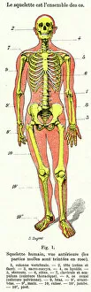
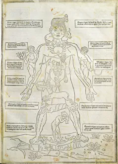
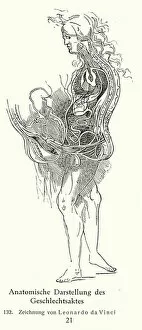
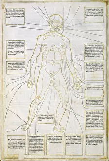
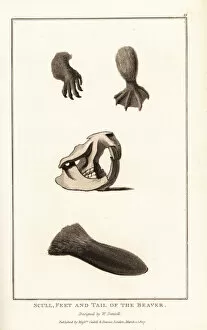
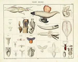
"Exploring the Intricacies of Human and Animal Anatomy: A Journey Through Anatomical Charts" Delve into the fascinating world of anatomy with a captivating collection of anatomical charts spanning centuries. From Bernardino de Montana De Monserrate's groundbreaking work on human anatomy to intricate woodcuts from 15th-century Venice, these illustrations offer a glimpse into our understanding of the human body throughout history. The main organs of the chest, stomach, and even the penis are meticulously depicted in engravings found in Libro de la anatomia del hombre. This Spanish doctor's comprehensive study sheds light on Charles V's era and provides valuable insights into medical knowledge during that time. A front view illustration from Fasciculus medicinae showcases a detailed human skeleton, allowing us to marvel at its complexity. The woodcut technique used in this piece adds an artistic touch while preserving scientific accuracy. Venturing beyond humans, we encounter depictions of horse anatomy dating back to 1792. Engravings reveal both the intestines and skeletal structure of these majestic creatures, offering invaluable information for veterinary medicine enthusiasts or simply those curious about equine biology. Continuing our exploration, aquatint prints unveil intriguing details about Eurasian beavers. Skulls, feet, and tails are meticulously portrayed with precision in this 1807 artwork—an excellent resource for zoologists studying these remarkable animals. Dive deep beneath the waves as lithographs from 1841 expose us to bivalve molluscs' inner workings. These intricate drawings provide an intimate look at their complex internal structures—a treasure trove for marine biologists seeking greater comprehension of these underwater organisms. Journeying further through lithograph illustrations from the same year reveals mesmerizing insights into jellyfish species' anatomy. Delicate yet resilient bodies come alive on paper as we examine their unique features—adding another layer to our understanding of marine life diversity. Step back in time once more with lithographs showcasing the anatomy of bivalve molluscs.

© Copyright Media Storehouse, All Rights Reserved 2003 - 2025
Facebook     Pinterest     100% Payment Secure     Reviews IO