
Allergic Reaction Collection
Allergic reactions can be triggered by various factors, ranging from insect stings to microscopic organisms lurking in our surroundings


All Professionally Made to Order for Quick Shipping
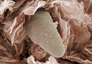
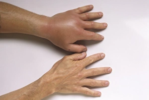
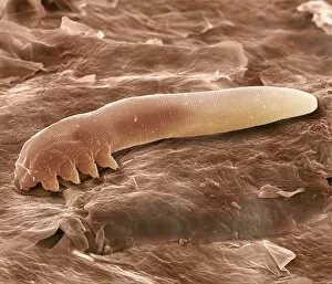
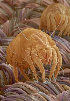
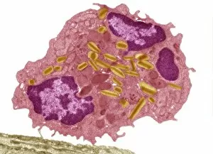
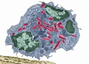
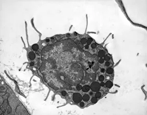
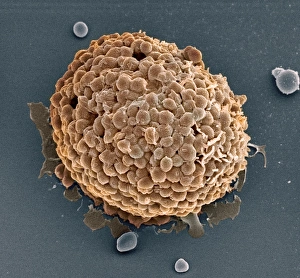
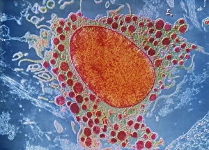
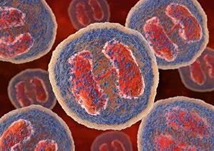
Allergic reactions can be triggered by various factors, ranging from insect stings to microscopic organisms lurking in our surroundings. One such example is the aftermath of a wasp sting, which can lead to an intense allergic reaction. The venom injected by the wasp causes swelling and redness around the affected area, often accompanied by pain and itching. On a much smaller scale, we delve into the world of eyelash mites. These tiny creatures reside on our eyelashes and feed off dead skin cells. However, their presence can sometimes cause discomfort for individuals with allergies. Under scanning electron microscopy (SEM), their tails become visible as they cling onto each lash. Moving on to dust mites - another common allergen found in households worldwide. SEM images reveal these minuscule arachnids up close; their translucent bodies covered in bristles that help them navigate through household dust particles. These microscopic pests are notorious for triggering allergic reactions such as sneezing, coughing, and itchy eyes. The intricate details captured under SEM expose the true nature of these allergens - whether it's a single dust mite or an entire colony thriving within your home's nooks and crannies. Their presence may go unnoticed but can wreak havoc on sensitive individuals who experience severe allergic responses upon exposure. Understanding the sources of allergic reactions is crucial for managing one's health effectively. From wasp stings to unseen inhabitants like dust mites or eyelash mites, being aware of potential triggers empowers us to take necessary precautions and seek appropriate treatments when needed.