
Air Sac Collection
In the vast expanse of Sublette County, Wyoming, amidst the breathtaking landscapes of the USA, a mesmerizing spectacle unfolds


All Professionally Made to Order for Quick Shipping
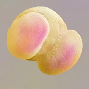
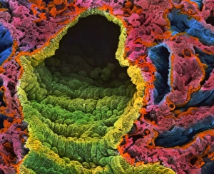
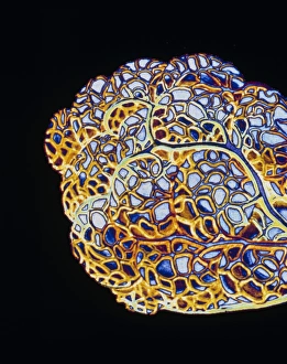
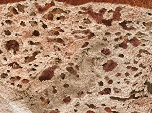
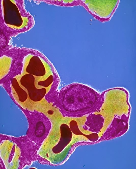
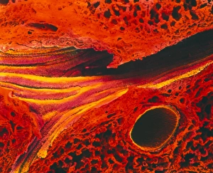
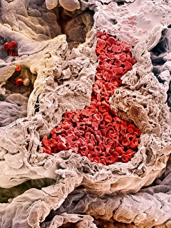
In the vast expanse of Sublette County, Wyoming, amidst the breathtaking landscapes of the USA, a mesmerizing spectacle unfolds. As Spring dawns upon us, a male Greater Sage Grouse takes center stage on a lek. With feathers puffed and wings spread wide, he displays his vibrant plumage in an attempt to attract a mate. Meanwhile, deep within our own bodies lies another marvel - the intricate network of air sacs known as lung alveoli. These tiny structures resemble delicate artwork under the microscope's lens (artwork C016 / 7680). Just like nature's creations outside our windows they can awe-inspiring in their complexity. But let us delve even further into this microscopic world. A phantom midge larva (C017 / 8348) dances through our imagination as we explore these lung alveoli up close. Their beauty is magnified by scanning electron microscopy (SEM), revealing intricate details that captivate our senses. Amongst these images emerges one of Scots pine pollen grain captured by SEM - a testament to how interconnected all living beings truly are. The exchange between plants and animals extends even to the tiniest corners of existence. Returning to our lungs' remarkable architecture, we find ourselves immersed in computer illustrations showcasing alveoli at work – expanding and contracting with each breath we take, and is here that life-giving oxygen enters our bloodstream while carbon dioxide exits through capillaries nestled within alveolar septa (F/col TEM). A colored SEM image brings it all together – displaying lungs adorned with countless bronchi and alveoli like jewels embedded within them. This masterpiece reminds us of both the fragility and resilience inherent in every breath we draw. So whether it be on Sublette County's grand stage or within the depths of your own being, let us celebrate these extraordinary air sacs – guardians of life itself – for they connect us to the very essence of existence.