
Adhesion Collection
"Exploring the Fascinating World of Adhesion: From Water Droplets to Cell Junctions" Spherical water droplets on leaf surface with high contact angle


All Professionally Made to Order for Quick Shipping
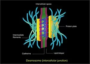
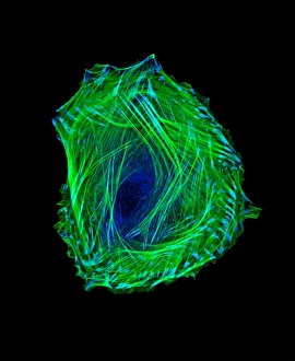
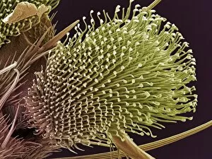
"Exploring the Fascinating World of Adhesion: From Water Droplets to Cell Junctions" Spherical water droplets on leaf surface with high contact angle: Nature's way of showcasing adhesion in action, as water forms beautiful beads on a leaf. Illustration of adhesions, thick and fibrous bands: Visualizing the intricate network of adhesive bonds that hold tissues together, like strong ropes connecting different parts. Cluster fly foot, SEM: Zooming in on a cluster fly's foot reveals tiny structures designed for maximum grip and adhesion to surfaces. Embryonic smooth muscle cell C018 / 8595: Witnessing the early stages of life through microscopic images, where cells begin forming connections essential for development and function. Bladder adhesions: Shedding light on a medical condition where abnormal tissue attachments cause discomfort and complications within the bladder. Migrating cell, illustration C018 / 0754: Journeying into the world of cellular movement, witnessing how cells adhere and migrate during crucial processes like wound healing or embryogenesis. Yellow dung fly's foot, SEM: Delving into nature's engineering marvels as we explore the specialized feet of yellow dung flies equipped with adhesive structures for walking upside down effortlessly. Pulvilli on a fly's foot, SEM (Scanning Electron Microscope): Unveiling the incredible adaptations found in insects' feet - pulvilli act like sticky pads enabling them to cling onto various surfaces with ease. Beetle foot, SEM (Scanning Electron Microscope): Examining beetles' remarkable ability to navigate their environment using specially evolved adhesive pads on their feet. Desmosome cell junction artwork: Appreciating art inspired by science as we admire an artistic representation capturing desmosomes – critical intercellular junctions ensuring strong adhesion between neighboring cells. In this captivating journey, we explore the diverse aspects.