Fraud Blocker Skip to main content

3 D Visualisation Collection

"Unlocking the Microscopic World

Background image3 D Visualisation Collection: Cucumber mosaic virus, computer model

Cucumber mosaic virus, computer model
Cucumber mosaic virus (CMV), computer model. This image was created using molecular modelling software and data from X-ray crystallography

Background image3 D Visualisation Collection: Murine norovirus with antibody fragments

Murine norovirus with antibody fragments
Murine norovirus (MNV) with antibody fragments, computer model. This image was created using molecular modelling software and data from cryo- electron microscopy

Background image3 D Visualisation Collection: Cucumber necrosis virus, computer model

Cucumber necrosis virus, computer model
Cucumber necrosis virus (CNV), computer model. This image was created using molecular modelling software and data from cryo-electron microscopy

Background image3 D Visualisation Collection: Human rhinovirus, computer model

Human rhinovirus, computer model
Human rhinovirus (HRV) with intercellular adhesion molecule 1 (ICAM-1), computer model. This image was created using UCSF Chimera molecular modelling software and data from cryo-electron microscopy

Background image3 D Visualisation Collection: Ribgrass mosaic virus, computer model

Ribgrass mosaic virus, computer model
Ribgrass mosaic virus (RMV), computer model. This image was created using UCSF Chimera molecular modelling software and fibre X-ray diffraction

Background image3 D Visualisation Collection: Human rhinovirus with antibodies

Human rhinovirus with antibodies
Human rhinovirus (HRV) with antibody fragments, computer model. This image was created using molecular modelling software and data from cryo- electron microscopy

Background image3 D Visualisation Collection: Cucumber mosaic virus with antibodies

Cucumber mosaic virus with antibodies
Cucumber mosaic virus (CMV) with antibody fragments attached, computer model. This image was created using molecular modelling software and data from cryo-electron microscopy

Background image3 D Visualisation Collection: Bacteriophage phi29, computer model

Bacteriophage phi29, computer model
Bacteriophage phi29. Cross section computer model of phi29 created using UCSF Chimera molecular modelling software and data from cryo-electron microscopy

Background image3 D Visualisation Collection: Murine norovirus, computer model

Murine norovirus, computer model
Murine norovirus (MNV), computer model. This image was created using molecular modelling software and data from cryo-electron microscopy

Background image3 D Visualisation Collection: Simian immunodeficiency virus (SIV)

Simian immunodeficiency virus (SIV), computer model. Cross section of SIV created using 3-D molecular modelling software and data from cryo- electron microscopy

Background image3 D Visualisation Collection: Sindbis virus, computer model

Sindbis virus, computer model
Sindbis virus. Computer model of sindbis virus created using UCSF Chimera molecular modelling software and data from cryo-electron microscopy. It shows the outer glycoprotein shell of the virus

Background image3 D Visualisation Collection: Semliki Forest virus, computer model

Semliki Forest virus, computer model
Semliki Forest virus (SFV), computer model. This image was created using UCSF Chimera molecular modelling software and data from cryo-electron microscopy

Background image3 D Visualisation Collection: Yeast cell, electron tomogram

Yeast cell, electron tomogram image. Yeast cell, Schizosaccharomyces pombe, created using a 3-D electron microscope. This involves firing beams of electrons from many different angles to create


All Professionally Made to Order for Quick Shipping

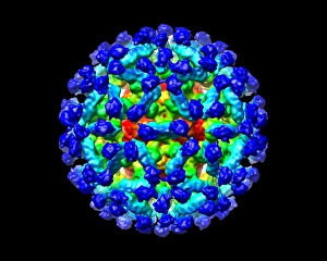
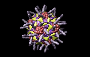
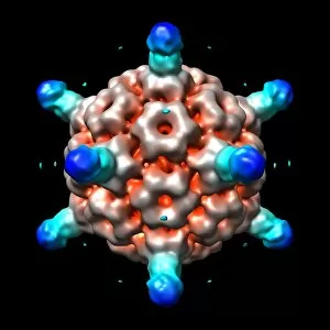
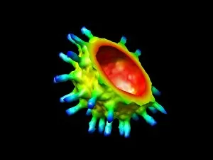
"Unlocking the Microscopic World: 3D Visualisation Reveals Intricate Details of Viruses and Antibodies" Step into the realm of cutting-edge scientific research as 3D visualisation takes us on a journey through the microscopic world. Using advanced computer models, scientists have delved into the intricate structures of various viruses and their interactions with antibodies. One such breakthrough is Bacteriophage phi29, where a computer model has unraveled its complex architecture. This virus, known for infecting bacteria, showcases its unique shape and components in stunning detail. Simian immunodeficiency virus (SIV), closely related to HIV, has also been brought to life through 3D visualisation. By mapping out its structure, researchers gain valuable insights into potential targets for antiviral therapies. In another fascinating study, Murine norovirus with antibody fragments reveals how these tiny proteins bind to specific regions of the virus. This knowledge paves the way for designing more effective treatments against this common cause of gastroenteritis. Continuing our exploration, we encounter Cucumber necrosis virus and Human rhinovirus – both meticulously recreated using computer models. These representations offer invaluable information about their structural intricacies that can aid in developing targeted interventions against plant diseases and respiratory infections respectively. The captivating Ribgrass mosaic virus comes next; its detailed computer model provides clues about how it interacts with host plants at a molecular level. Understanding these mechanisms could potentially lead to strategies for crop protection against viral pathogens. Human rhinovirus returns but this time accompanied by antibodies - an exciting development in combating common colds. The interaction between these two entities is now better understood thanks to 3D visualisation techniques, opening doors towards novel therapeutic approaches. Bacteriophage P22 follows suit as another example where computational modelling sheds light on its inner workings. With further investigation into this bacterial predator's structure and function, new avenues may emerge for combating antibiotic-resistant bacteria.