Jigsaw Puzzle : Diagram of the bones of the right leg and hip


![]()
Jigsaw Puzzles from Mary Evans Picture Library
Diagram of the bones of the right leg and hip
Diagram of the bones of the right leg, showing the joint with the pelvis at the hip
Mary Evans Picture Library makes available wonderful images created for people to enjoy over the centuries
Media ID 4356360
© Mary Evans Picture Library 2015 - https://copyrighthub.org/s0/hub1/creation/maryevans/MaryEvansPictureID/10081838
Anatomical Ankle Bone Bones Diagram Femur Fibula Foot Human Joint Knee Legs Limb Lower Patella Pelvis Shin Skeleton Tarsus Tibia Toes Metatarsus Phalanges
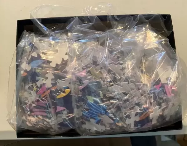
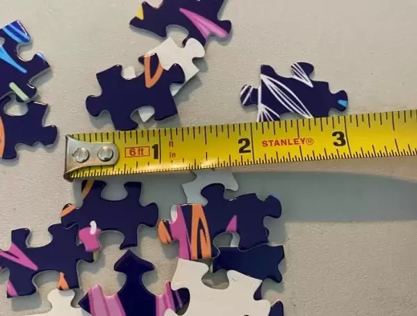
Jigsaw Puzzle (252 Pieces)
Discover the intricacies of the human body with our educational and captivating Media Storehouse Jigsaw Puzzles. This particular puzzle features a detailed diagram of the bones in the right leg and hip joint, sourced from Mary Evans Prints Online. Ideal for anatomy enthusiasts, students, or anyone with a curiosity for the intricacies of the human body, this puzzle offers a unique and engaging way to learn. Assemble the pieces to reveal the complex relationship between the bones in your right leg and hip, providing a tangible understanding of this essential joint. Puzzle pieces are made from high-quality materials, ensuring a satisfying puzzle experience. Join us in exploring the wonders of the human body, one puzzle at a time.
Made in the USA, 252-piece puzzles measure 11" x 14" (27.9 x 35.5 cm). Every puzzle is meticulously printed on glossy photo paper, which has a strong 1.33 mm thickness. Delivered in a black storage cardboard box, these puzzles are both stylish and practical. (Note: puzzles contain small parts and are not suitable for children under 3 years of age.)
Jigsaw Puzzles are an ideal gift for any occasion
Estimated Product Size is 28cm x 35.6cm (11" x 14")
These are individually made so all sizes are approximate
Artwork printed orientated as per the preview above, with portrait (vertical) orientation to match the source image.
EDITORS COMMENTS
This fascinating historical print from 1908 showcases a detailed diagram of the bones of the right leg and hip, providing a glimpse into the intricate anatomy of the human body. The illustration highlights key components such as the femur, tibia, fibula, patella, and metatarsus, offering valuable insight into the skeletal structure of this crucial part of our bodies.
The joint with the pelvis at the hip is also prominently featured in this diagram, emphasizing how these bones work together to support movement and stability. From the ankle to the knee to the toes, every element is meticulously labeled and depicted with precision, making it an invaluable resource for medical professionals and students alike.
As we admire this image, we are reminded of the complexity and sophistication of our own bodies. The history behind this anatomical illustration adds another layer of intrigue, serving as a testament to centuries of study and research dedicated to understanding human physiology.
Whether you have a passion for anatomy or simply appreciate fine artistry, this print offers a unique blend of scientific accuracy and visual appeal. It serves as a timeless reminder of our shared humanity and the remarkable intricacies that make up our physical selves.
MADE IN THE USA
Safe Shipping with 30 Day Money Back Guarantee
FREE PERSONALISATION*
We are proud to offer a range of customisation features including Personalised Captions, Color Filters and Picture Zoom Tools
SECURE PAYMENTS
We happily accept a wide range of payment options so you can pay for the things you need in the way that is most convenient for you
* Options may vary by product and licensing agreement. Zoomed Pictures can be adjusted in the Cart.