Jigsaw Puzzle : Anatomy in the 18th C


![]()
Jigsaw Puzzles from Mary Evans Picture Library
Anatomy in the 18th C
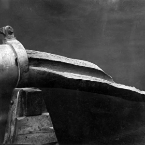
a skull, teeth, the interior of a knee (below left) some marrow and a mucous gland from the humerus (below right) Date: Circa 1760
Mary Evans Picture Library makes available wonderful images created for people to enjoy over the centuries
Media ID 7155127
© Mary Evans Picture Library 2015 - https://copyrighthub.org/s0/hub1/creation/maryevans/MaryEvansPictureID/10158175
1760 Gland Humerus Knee Marrow Skull Teeth Mucous
Jigsaw Puzzle (252 Pieces)
Discover the fascinating world of 18th century anatomy with our exquisite jigsaw puzzle from Media Storehouse, featuring an intricately detailed illustration from Mary Evans Prints Online. Delve into this captivating piece of history and assemble the intricately depicted bones and organs, including a skull, teeth, the interior of a knee, and even a mucous gland from the humerus. Dating back to circa 1760, this puzzle offers a unique and educational experience for puzzle enthusiasts and history buffs alike. Engage your mind and bring the past to life with this captivating anatomy puzzle from Media Storehouse.
Made in the USA, 252-piece puzzles measure 11" x 14" (27.9 x 35.5 cm). Every puzzle is meticulously printed on glossy photo paper, which has a strong 1.33 mm thickness. Delivered in a black storage cardboard box, these puzzles are both stylish and practical. (Note: puzzles contain small parts and are not suitable for children under 3 years of age.)
Jigsaw Puzzles are an ideal gift for any occasion
Estimated Product Size is 28cm x 35.6cm (11" x 14")
These are individually made so all sizes are approximate
Artwork printed orientated as per the preview above, with portrait (vertical) orientation to match the source image.
EDITORS COMMENTS
This print, dated circa 1760, showcases an intriguing collection of human anatomical specimens from the 18th century. The image, which is believed to have been produced around the same period as William Hunter's influential anatomy textbooks, offers a glimpse into the scientific curiosity and exploration of the human body during this time. On the left side of the print, an elegant skull and a row of teeth are displayed, their intricate details revealing the artist's meticulous attention to anatomical accuracy. The interior of a knee joint, situated below the skull, is depicted in remarkable detail, with the meniscus, patella, and other structures clearly visible. The artist's skillful rendering of the complex knee anatomy was essential for the advancement of surgical techniques during this era. Beneath the knee, two smaller illustrations capture the intrigue of lesser-known anatomical structures. The first, a cross-section of a humerus bone, reveals the presence of a mucous gland. This gland, which produces synovial fluid to lubricate the joint, was a subject of fascination for medical researchers of the time. The second illustration, nestled next to it, displays a section of marrow from the same bone. The study of bone marrow was crucial in understanding the role it plays in the production of blood cells, contributing significantly to the field of hematology. This print, with its intricate details and scientific accuracy, offers a unique window into the world of 18th-century anatomical exploration and discovery. Its preservation allows us to appreciate the depth of knowledge and curiosity that drove medical advancements during this period.
MADE IN THE USA
Safe Shipping with 30 Day Money Back Guarantee
FREE PERSONALISATION*
We are proud to offer a range of customisation features including Personalised Captions, Color Filters and Picture Zoom Tools
FREE COLORIZATION SERVICE
You can choose advanced AI Colorization for this picture at no extra charge!
SECURE PAYMENTS
We happily accept a wide range of payment options so you can pay for the things you need in the way that is most convenient for you
* Options may vary by product and licensing agreement. Zoomed Pictures can be adjusted in the Cart.