Fraud Blocker Skip to main content

Home > All Images > 2012 > February > 21 Feb 2012

Images Dated 21st February 2012 (page 7)

387 items

Background imageImages Dated 21st February 2012: German Foot Soldier 1515

German Foot Soldier 1515
German foot soldiers Date: circa 1515

Background imageImages Dated 21st February 2012: C18 Austrian Military

C18 Austrian Military
Austrian Soldiers Date: 1728

Background imageImages Dated 21st February 2012: Folklore / Insects

Folklore / Insects
CROSSEs ACARI Insect which Crosse believed he had created spontaneously and inadvertently in the course of electro-chemical experiments Date: 1837

Background imageImages Dated 21st February 2012: Paramecium protozoan, light micrograph

Paramecium protozoan, light micrograph
Paramecium protozoan. Differential interference contrast micrograph of a Paramecium sp. protozoan. This ciliate protozoan inhabits freshwater, where it feeds mainly on bacteria

Background imageImages Dated 21st February 2012: Glycine crystals, light micrograph

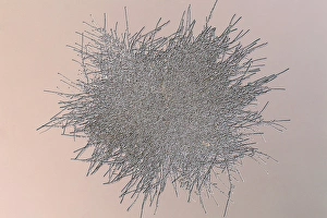
Glycine crystals, light micrograph
Glycine crystals. Polarised light micrograph of crystals of glycine, the simplest of the amino acids. Glycine consists of a central carbon atom, to which are attached two hydrogen atoms

Background imageImages Dated 21st February 2012: American oak leaf, light micrograph

American oak leaf, light micrograph
American oak leaf. Light micrograph showing the veins in a leaf from an American oak (Quercus rubra) tree. Magnification: x60 when printed at 10 centimetres wide

Background imageImages Dated 21st February 2012: Fluke worm, light micrograph

Fluke worm, light micrograph
Fluke worm. Light micrograph of a section through a parasitic trematode (fluke) worm. These flatworms often parasitise birds and other animals but can infect humans

Background imageImages Dated 21st February 2012: American oak leaf, light micrograph

American oak leaf, light micrograph
American oak leaf. Light micrograph showing the veins in a leaf from an American oak (Quercus rubra) tree. Magnification: x12 when printed at 10 centimetres wide

Background imageImages Dated 21st February 2012: Potato starch grains, light micrograph

Potato starch grains, light micrograph
Potato starch grains. Polarised light micrograph of starch grains from a potato. Magnification: x120 when printed 10 centimetres wide

Background imageImages Dated 21st February 2012: Potato starch grains, light micrograph

Potato starch grains, light micrograph
Potato starch grains. Polarised light micrograph of starch grains from a potato. Magnification: x120 when printed 10 centimetres wide

Background imageImages Dated 21st February 2012: Wild pansy (Viola tricolor) flower

Wild pansy (Viola tricolor) flower. Close-up of a flower from a Viola tricolor plant, showing the papillae (finger-like) and petal structure

Background imageImages Dated 21st February 2012: Butterfly wing scales

Butterfly wing scales. Close-up of scales on the wing of a blue morpho (Morpho peleides) butterfly. Magnification: x12 when printed at 10 centimetres wide

Background imageImages Dated 21st February 2012: Swan feather, light micrograph

Swan feather, light micrograph
Swan feather. Differential interference contrast micrograph showing the surface structure of a feather from a mute swan (Cygnus olor). Magnification: x120 when printed at 10 centimetres wide

Background imageImages Dated 21st February 2012: Glycine crystals, light micrograph

Glycine crystals, light micrograph
Glycine crystals. Polarised light micrograph of crystals of glycine, the simplest of the amino acids. Glycine consists of a central carbon atom, to which are attached two hydrogen atoms

Background imageImages Dated 21st February 2012: Potato starch grains, light micrograph

Potato starch grains, light micrograph
Potato starch grains. Polarised light micrograph of starch grains from a potato. Magnification: x120 when printed 10 centimetres wide

Background imageImages Dated 21st February 2012: Ciprofloxacin crystals, light micrograph

Ciprofloxacin crystals, light micrograph
Ciprofloxacin crystals. Fluorescence light micrograph of crystals of the broad-spectrum antibiotic drug ciprofloxacin. Magnification: x120 when printed 10 centimetres wide

Background imageImages Dated 21st February 2012: Mosquito wing, light micrograph

Mosquito wing, light micrograph
Mosquito wing. Light micrograph showing the structure of a wing from a Culex pipiens mosquito. Magnification: x120 when printed 10 centimetres wide

Background imageImages Dated 21st February 2012: Fluke worm, light micrograph

Fluke worm, light micrograph
Fluke worm. Light micrograph of a section through an Echinostoma revolutum parasitic trematode (fluke) worm. These flatworms often parasitise birds but can infect humans

Background imageImages Dated 21st February 2012: Spirogyra algae, light micrograph

Spirogyra algae, light micrograph
Spirogyra algae. Polarised light micrograph of Spirogyra sp. algae. This filamentous green algae is named for the spiral arrangement of its chloroplasts (green)

Background imageImages Dated 21st February 2012: Mouse flea, light micrograph

Mouse flea, light micrograph
Mouse flea. Light micrograph of a Megabothris turbidus flea. These fleas live mainly on rodents. Magnification: x20 when printed 10 centimetres wide

Background imageImages Dated 21st February 2012: Spirogyra algae, light micrograph

Spirogyra algae, light micrograph
Spirogyra algae. Light micrograph of Spirogyra sp. algae. This filamentous green algae is named for the spiral arrangement of its chloroplasts (green)

Background imageImages Dated 21st February 2012: Ciprofloxacin crystals, light micrograph

Ciprofloxacin crystals, light micrograph
Ciprofloxacin crystals. Fluorescence light micrograph of crystals of the broad-spectrum antibiotic drug ciprofloxacin. Magnification: x120 when printed 10 centimetres wide

Background imageImages Dated 21st February 2012: Pulpi geode, Spain

Pulpi geode, Spain
MODEL RELEASED. Pulpi geode, Almeria, Spain. Geologist inside the Pulpi geode, the largest geode ever discovered. Geodes are rocks that are hollow in the centre

Background imageImages Dated 21st February 2012: Butterfly wing eye spot

Butterfly wing eye spot. Close-up of an eye spot on the wing of a blue morpho (Morpho peleides) butterfly. Magnification: x12 when printed at 10 centimetres wide

Background imageImages Dated 21st February 2012: Mouse flea, light micrograph

Mouse flea, light micrograph
Mouse flea. Light micrograph of a Megabothris turbidus flea. These fleas live mainly on rodents. Magnification: x20 when printed 10 centimetres wide

Background imageImages Dated 21st February 2012: Epistylis protozoan colony on algae

Epistylis protozoan colony on algae. Light micrograph of a colony of Epistylis sp. protozoa (centre right) attached to a green algae filament (lower left)

Background imageImages Dated 21st February 2012: Fluke worm, light micrograph

Fluke worm, light micrograph
Fluke worm. Light micrograph of a section through the head of an Echinostoma revolutum parasitic trematode (fluke) worm. These flatworms often parasitise birds but can infect humans

Background imageImages Dated 21st February 2012: Pulpi geode, Spain

Pulpi geode, Spain
MODEL RELEASED. Pulpi geode, Almeria, Spain. Geologist inside the Pulpi geode, the largest geode ever discovered. Geodes are rocks that are hollow in the centre

Background imageImages Dated 21st February 2012: Varroa jacobsoni mite, light micrograph

Varroa jacobsoni mite, light micrograph
Varroa jacobsoni mite. Differential interference contrast micrograph of a Varroa jacobsoni honey bee mite. This arachnid is a relatively harmless parasite of Asian honey bees (Apis cerana)

Background imageImages Dated 21st February 2012: Heartworms, light micrograph

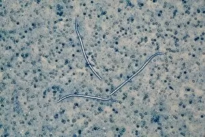
Heartworms, light micrograph
Heartworms. Light micrograph of three Dirofilaria immitis heartworms. During the adult stage of their life cycle, these nematode worms live in the right ventricle of animal hearts

Background imageImages Dated 21st February 2012: Tapeworm, light micrograph

Tapeworm, light micrograph
Tapeworm. Light micrograph of a section through a Taenia taeniaeformis parasitic tapeworm, showing the hooks around its mouth (upper right), which it uses to cling to the intestinal wall of its host

Background imageImages Dated 21st February 2012: Glycine crystals, light micrograph

Glycine crystals, light micrograph
Glycine crystals. Polarised light micrograph of crystals of glycine, the simplest of the amino acids. Glycine consists of a central carbon atom, to which are attached two hydrogen atoms

Background imageImages Dated 21st February 2012: Green tiger beetle wing

Green tiger beetle wing
Green tiger beetle. Close-up of the wing of a Green tiger beetle (Cicindela campestris), showing the surface pits that refract and reflect light to give an iridescent appearance

Background imageImages Dated 21st February 2012: Parasitic flatworm, light micrograph

Parasitic flatworm, light micrograph
Parasitic flatworm. Light micrograph of a section through a Diplozoon paradoxum parasitic monogenean worm. Monogenea are very small parasitic flatworms mainly found on skin or gills of fish

Background imageImages Dated 21st February 2012: Swan feather, light micrograph

Swan feather, light micrograph
Swan feather. Differential interference contrast micrograph showing the surface structure of a feather from a mute swan (Cygnus olor). Magnification: x12 when printed at 10 centimetres wide

Background imageImages Dated 21st February 2012: Penicillium mould colony

Penicillium mould colony. Penicillium is used to produce the antibiotic penicillin. Magnification: x60 when printed 10 centimetres wide

Background imageImages Dated 21st February 2012: Proteus amoeba, light micrograph

Proteus amoeba, light micrograph
Proteus amoeba. Differential interference contrast micrograph of an Amoeba proteus protozoan. This freshwater single-celled organism feeds on bacteria and smaller protozoa

Background imageImages Dated 21st February 2012: Swan feather, light micrograph

Swan feather, light micrograph
Swan feather. Differential interference contrast micrograph showing the surface structure of a feather from a mute swan (Cygnus olor). Magnification: x120 when printed at 10 centimetres wide

Background imageImages Dated 21st February 2012: Butterfly wing scales

Butterfly wing scales. Light micrograph of scales on the wing of a blue morpho (Morpho peleides) butterfly. Magnification: x60 when printed at 10 centimetres wide

Background imageImages Dated 21st February 2012: Threadworm, light micrograph

Threadworm, light micrograph
Threadworm. Light micrograph of a female Enterobius vermicularis threadworm. Threadworms are nematode worms that parasitise the large intestine and caecum of many animals

Background imageImages Dated 21st February 2012: Tick, light micrograph

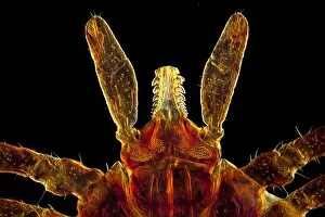
Tick, light micrograph
Tick. Light micrograph of the head of a male castor bean tick (Ixodes ricinus) showing its mouthparts (yellow). The mouthparts of a tick consist of three visible components

Background imageImages Dated 21st February 2012: Pulpi geode, Spain

Pulpi geode, Spain
MODEL RELEASED. Pulpi geode, Almeria, Spain. Geologist inside the Pulpi geode, the largest geode ever discovered. Geodes are rocks that are hollow in the centre

Background imageImages Dated 21st February 2012: Pulpi geode, Spain

Pulpi geode, Spain
Pulpi geode, Almeria, Spain. Inside of the Pulpi geode, the largest geode ever discovered. Geodes are rocks that are hollow in the centre. The interior is often lined with crystals

Background imageImages Dated 21st February 2012: Tick, light micrograph

Tick, light micrograph
Tick. Light micrograph of the head of a male castor bean tick (Ixodes ricinus) showing its mouthparts (yellow). The mouthparts of a tick consist of three visible components

Background imageImages Dated 21st February 2012: Glycine crystals, light micrograph

Glycine crystals, light micrograph
Glycine crystals. Polarised light micrograph of crystals of glycine, the simplest of the amino acids. Glycine consists of a central carbon atom, to which are attached two hydrogen atoms

Background imageImages Dated 21st February 2012: Mouse flea, light micrograph

Mouse flea, light micrograph
Mouse flea. Light micrograph of the head of a Megabothris turbidus flea. These fleas live mainly on rodents. Magnification: x120 when printed 10 centimetres wide

Background imageImages Dated 21st February 2012: Fluke worm, light micrograph

Fluke worm, light micrograph
Fluke worm. Light micrograph of a section through a parasitic trematode (fluke) worm. These flatworms often parasitise birds and other animals but can infect humans

Background imageImages Dated 21st February 2012: Spirogyra algae, light micrograph

Spirogyra algae, light micrograph
Spirogyra algae. Polarised light micrograph of Spirogyra sp. algae. This filamentous green algae is named for the spiral arrangement of its chloroplasts (green)


All Professionally Made to Order for Quick Shipping


-

Why Choose Us?

We are a leading provider of Art Prints and Photo Gifts since 2004, working in partnership with a range of Sporting Clubs, Charities, Museums and Picture Libraries. A large share of profits from any of their images will go directly towards supporting that charity or club. Our archive of images is carefully curated to bring you a wide range of subjects, including landscapes, wildlife, architecture, and more. We ship from our partner labs in the UK, USA, EU (Netherlands) and Australia.
+

How do I place an order?

Ordering is quick & easy - Just follow 5 Simple Steps:
  1. Find your image: Use our search box or browse our online photo Collections to find the image you want.

  2. Choose your print format: Select your desired product and add it to your cart.

  3. Enter your details: If you're a returning customer, simply enter your email address and password, and we'll fill in your billing and shipping address details. All personal details are held securely and are fully GDPR compliant. As standard, we remove all Personally Identifiable Information after 12 months.

  4. Pay for your purchase: We use state-of-the-art security for online shopping and do not have access to your card details.

  5. Sit back and relax: We'll email you confirmation of your order and when it's dispatched. Registered customers can also track orders in the 'My Account' area.

+

How do I pay for an order?

You can pay for your order with most of the major credit and debit cards, or PayPal. For added security, major financial institutions process payment details separately and securely on our behalf. We do not have access to your online payment card details. Online payments are preferred however we do take phone orders. For UK customers only we accept cheques issued against a UK bank.
+

Is my data safe?

We take data security very seriously. We do not have access to your full card details and all payments are requested over a fully secure connection. Additionally, we fully comply with current European and GDPR legislation, and automatically remove all personal data after at most 12 months (unless you have an account with us and have signed in recently). We also have a strict opt-in policy and would never sell your personal details. Your data is only used to fulfill your order promptly and efficiently.
+

Is wall art ready to hang?

For quick and easy installation all wall art, including framed prints, canvas prints and metal prints are supplied with a ready to hang solution on the back. Generally, saw tooth hangers are applied as they allow wall art to hang flush against the wall. The serrated edge of the hanger prevents the frame from shifting or tilting when hung.
+

Are the photo prints fade resistant?

Yes, we use archival quality photo paper photographic paper for vivid reproduction Prints are an accurate representations of the original artwork, which is preserved for artistic character and authenticity. We guarantee they match previews shown on our web site