Fraud Blocker Skip to main content

Home > All Images > 2012 > February > 1 Feb 2012

Images Dated 1st February 2012 (page 6)

330 items

Background imageImages Dated 1st February 2012: London: tower Bridge

London: tower Bridge
LONDON:TOWER BRIDGE

Background imageImages Dated 1st February 2012: THE GREAT SPHINX The Great Sphinx at Giza, Egypt. 4th Dynasty

THE GREAT SPHINX The Great Sphinx at Giza, Egypt. 4th Dynasty

Background imageImages Dated 1st February 2012: DANUBE: STEAMBOAT. Crew members swabbing the deck of a steamboat on the Danube River at sunrise

DANUBE: STEAMBOAT. Crew members swabbing the deck of a steamboat on the Danube River at sunrise, as it departs Bratislava, Slovakia. Photographed c1974

Background imageImages Dated 1st February 2012: MINNESOTA: LAKE ITASCA. A view of the headwaters of the Mississippi River at Lake Itasca

MINNESOTA: LAKE ITASCA. A view of the headwaters of the Mississippi River at Lake Itasca, Minnesota, at sunrise. Photographed c1974

Background imageImages Dated 1st February 2012: BIERSTADT: MATTERHORN. Sunrise on the Matterhorn. Oil on canvas by Albert Bierstadt, 19th century

BIERSTADT: MATTERHORN. Sunrise on the Matterhorn. Oil on canvas by Albert Bierstadt, 19th century

Background imageImages Dated 1st February 2012: MISSISSIPPI RIVER: BLUFFS. Bluffs overlooking the Mississippi River

MISSISSIPPI RIVER: BLUFFS. Bluffs overlooking the Mississippi River, viewed at sunrise from Riverview Park in Hannibal, Missouri. Photographed c1974

Background imageImages Dated 1st February 2012: PALMER: MISSISSIPPI, c1862. The Mississippi in Time of Peace

PALMER: MISSISSIPPI, c1862. The Mississippi in Time of Peace. Pencil, watercolor and gouache drawing by Fanny Palmer, c1862

Background imageImages Dated 1st February 2012: COCONUT PALMS (Cocos nucifer) at sunset. Gilbert Islands, Micronesia

COCONUT PALMS (Cocos nucifer) at sunset. Gilbert Islands, Micronesia

Background imageImages Dated 1st February 2012: EGYPT: NILE SCENE. Sunset over the Nile River in Nubia, Egypt. Photographed c1970

EGYPT: NILE SCENE. Sunset over the Nile River in Nubia, Egypt. Photographed c1970

Background imageImages Dated 1st February 2012: AUSTRIA: DANUBE. A view of the Danube River near Vienna, Austria. Photographed c1974

AUSTRIA: DANUBE. A view of the Danube River near Vienna, Austria. Photographed c1974

Background imageImages Dated 1st February 2012: MISSISSIPPI RIVER: SUNRISE. Waters of the Mississippi River reaching flood level at sunrise

MISSISSIPPI RIVER: SUNRISE. Waters of the Mississippi River reaching flood level at sunrise, near Louisiana, Missouri (Pike County). Photographed c1974

Background imageImages Dated 1st February 2012: MISSISSIPPI RIVER: SUNRISE. Sunrise over the Mississippi River, Hannibal, Missouri

MISSISSIPPI RIVER: SUNRISE. Sunrise over the Mississippi River, Hannibal, Missouri. Photographed c1974

Background imageImages Dated 1st February 2012: HOLY LAND: DEAD SEA. A sunset over the coast of the Dead Sea. Photograph, c1937

HOLY LAND: DEAD SEA. A sunset over the coast of the Dead Sea. Photograph, c1937

Background imageImages Dated 1st February 2012: IRAQ: SUNSET, 1932. Sunset along a river in Iraq. Photograph, 1932

IRAQ: SUNSET, 1932. Sunset along a river in Iraq. Photograph, 1932

Background imageImages Dated 1st February 2012: MISSISSIPPI RIVER: FISHING. A man seated between fishing poles beside the Mississippi River in St

MISSISSIPPI RIVER: FISHING. A man seated between fishing poles beside the Mississippi River in St. Louis, Missouri, near sunset. Photographed c1974

Background imageImages Dated 1st February 2012: IRAQ: RIVERBOAT, 1932. Riverboat photographed during sunset on a river in Iraq, 1932

IRAQ: RIVERBOAT, 1932. Riverboat photographed during sunset on a river in Iraq, 1932

Background imageImages Dated 1st February 2012: Grey mould fungus, light micrograph

Grey mould fungus, light micrograph
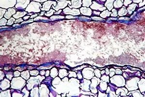
Grey mould fungus. Light micrograph of a section through onion tissue infected with grey mould fungus (Botrytis allii). Botrytis is a parasitic saprophyte mould that grows on wounded plants

Background imageImages Dated 1st February 2012: Female pine cone seed, light micrograph

Female pine cone seed, light micrograph
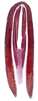
Female pine cone seed. Light micrograph of a section through a seed from a female pine (Pinus sp.) cone, showing the mature embryo and endosperm

Background imageImages Dated 1st February 2012: Calcareous phytoplankton, SEM

Calcareous phytoplankton, SEM
Calcareous phytoplankton. Scanning electron micrograph (SEM) of Emiliana. huxleyi. This small algal organism (coccolithophore) is surrounded by a skeleton (coccosphere)

Background imageImages Dated 1st February 2012: Pine stem, light micrograph

Pine stem, light micrograph
Pine stem. Light micrograph of a section through the stem of a pine (Pinus sp.) tree, showing a resin duct (lacuna, large, centre) in xylem tissue. The surrounding tissue is parenchyma cells (red)

Background imageImages Dated 1st February 2012: Pine stem, light micrograph

Pine stem, light micrograph
Pine stem. Light micrograph of a section through the stem of a pine (Pinus sp.) tree, showing tracheid cells (white-brown) in the xylem tissue

Background imageImages Dated 1st February 2012: Pine stem, light micrograph

Pine stem, light micrograph
Pine stem. Light micrograph of a section through the stem of a pine (Pinus sp.) tree, showing xylem tissue near the central pith. The xylem is made up of tracheid cells (light pink)

Background imageImages Dated 1st February 2012: Pear rust fungus, light micrograph

Pear rust fungus, light micrograph
Pear rust fungus. Light micrograph of a section through tissue from a juniper tree infected with pear rust fungus (Gymnosporangium sabinae, lower right)

Background imageImages Dated 1st February 2012: Male pine cone, light micrograph

Male pine cone, light micrograph
Male pine cone. Light micrograph of a section through a male pine (Pinus sp.) cone, showing microsporophylls (leaves, purple) bearing microsporangia (pink)

Background imageImages Dated 1st February 2012: Grey mould fungus, light micrograph

Grey mould fungus, light micrograph
Grey mould fungus. Light micrograph of a section through onion tissue infected with grey mould fungus (Botrytis allii). Botrytis is a parasitic saprophyte mould that grows on wounded plants

Background imageImages Dated 1st February 2012: Pine stem, light micrograph

Pine stem, light micrograph
Pine stem. Polarised light micrograph of a section through part of the stem of a pine (Pinus sp.) tree, showing the growth rings

Background imageImages Dated 1st February 2012: Pine stem, light micrograph

Pine stem, light micrograph
Pine stem. Light micrograph of a section through a single annual growth ring in the stem of a pine (Pinus sp.) tree, showing large thin-walled tracheid cells (whitish pink)

Background imageImages Dated 1st February 2012: Corn smut infection, light micrograph

Corn smut infection, light micrograph
Corn smut infection. Light micrograph of a section through tissue from a corn (Zea mays) plant infected with corn smut fungus (Ustilago maydis)

Background imageImages Dated 1st February 2012: Bramble rust fungus spores

Bramble rust fungus spores. Light micrograph of teliospores from the bramble rust fungus (Phragmidium violaceum). Magnification: x37 when printed 10 centimetres wide

Background imageImages Dated 1st February 2012: Rose mildew fungus, light micrograph

Rose mildew fungus, light micrograph
Rose mildew fungus. Light micrograph of a section through tissue from a rose plant infected with rose mildew fungus (Erysiphe pannosa)

Background imageImages Dated 1st February 2012: Pine stem, light micrograph

Pine stem, light micrograph
Pine stem. Light micrograph of a section through a single annual growth ring in the stem of a pine (Pinus sp.) tree, showing large thin-walled tracheid cells (light pink)

Background imageImages Dated 1st February 2012: Pine root, light micrograph

Pine root, light micrograph
Pine root. Light micrograph of a section through the root of a pine (Pinus sp.) tree. From outer to inwards: outer layer of peridium (dark red); cortex - made up of parenchyma cells (red)

Background imageImages Dated 1st February 2012: Colour temperature spectrum

Colour temperature spectrum. This spectrum shows how the overall hue of a source of visible light can vary in terms of a calculated characteristic known as colour temperature

Background imageImages Dated 1st February 2012: Female pine cone scale, light micrograph

Female pine cone scale, light micrograph
Female pine cone scale. Light micrograph of a section through a scale from a young ovulate female pine (Pinus sp.) cone, bearing an ovule (upper centre)

Background imageImages Dated 1st February 2012: Hollyhock rust fungus in a leaf

Hollyhock rust fungus in a leaf. Light micrograph of a section through a hollyhock (Alcea sp.) leaf infected with the parasitic Puccinia malvacearum rust, showing the teliospores (spores, brown-red)

Background imageImages Dated 1st February 2012: Pear scab infection, light micrograph

Pear scab infection, light micrograph
Pear scab infection. Light micrograph of a section through a pear (Pyrus sp.) infected with the Venturia pirinum fungus. This fungus causes scab, or black spot, of the pear fruit

Background imageImages Dated 1st February 2012: Black scab infection, light micrograph

Black scab infection, light micrograph
Black scab infection. Light micrograph of a section through a potato infected with black scab (Synchytrium endobioticum) fungus

Background imageImages Dated 1st February 2012: White rust fungus on a leaf

White rust fungus on a leaf. Light micrograph of a section through a plant leaf infected with white rust fungus (Albugo candida)

Background imageImages Dated 1st February 2012: Black scab infection, light micrograph

Black scab infection, light micrograph
Black scab infection. Light micrograph of a section through a potato infected with black scab (Synchytrium endobioticum) fungus

Background imageImages Dated 1st February 2012: Pine stem, light micrograph

Pine stem, light micrograph
Pine stem. Light micrograph of a section through the stem of a pine (Pinus sp.) tree, showing tracheid cells (white-brown) in the xylem tissue

Background imageImages Dated 1st February 2012: Female pine cone scale, light micrograph

Female pine cone scale, light micrograph
Female pine cone scale. Light micrograph of a section through a scale from a young ovulate female Scots pine (Pinus sylvestris) cone, bearing an ovule (upper right)

Background imageImages Dated 1st February 2012: Pine stem, light micrograph

Pine stem, light micrograph
Pine stem. Polarised light micrograph of a section through a single annual growth ring in the stem of a pine (Pinus sp.) tree, showing xylem tissue

Background imageImages Dated 1st February 2012: Powdery scab infection, light micrograph

Powdery scab infection, light micrograph
Powdery scab infection. Light micrograph of a section through a potato infected with powdery scab (Spongospora subterranea), showing pustules and spores. S

Background imageImages Dated 1st February 2012: Male pine cone, light micrograph

Male pine cone, light micrograph
Male pine cone. Light micrograph of a section through a male pine (Pinus sp.) cone, showing two microsporangia (pink) that contain numerous microspore pollen grains (blue)

Background imageImages Dated 1st February 2012: Rust fungus in a leaf, light micrograph

Rust fungus in a leaf, light micrograph
Rust fungus in a leaf. Light micrograph of a section through a barberry (Berberis sp.) leaf infected with Puccinia sp. rust, showing spore-bearing cluster-cups (cup-shaped)

Background imageImages Dated 1st February 2012: Rust fungus in a leaf, light micrograph

Rust fungus in a leaf, light micrograph
Rust fungus in a leaf. Light micrograph of a section through a wheat leaf infected with the parasitic Puccinia graminis rust

Background imageImages Dated 1st February 2012: Pear rust fungus, light micrograph

Pear rust fungus, light micrograph
Pear rust fungus. Light micrograph of a section through tissue from a juniper tree infected with pear rust fungus (Gymnosporangium sabinae)

Background imageImages Dated 1st February 2012: Plum pocket infection, light micrograph

Plum pocket infection, light micrograph
Plum pocket infection. Light micrograph of a section through a plum gall caused by the plum pocket fungus (Taphrina pruni)


All Professionally Made to Order for Quick Shipping


-

Why Choose Us?

We are a leading provider of Art Prints and Photo Gifts since 2003, working in partnership with a range of Sporting Clubs, Charities, Museums and Picture Libraries. A large share of profits from any of their images will go directly towards supporting that charity or club. Our archive of images is carefully curated to bring you a wide range of subjects, including landscapes, wildlife, architecture, and more. We ship from our partner labs in the UK, USA, EU (Netherlands) and Australia.
+

How do I place an order?

Ordering is quick & easy - Just follow 5 Simple Steps:
  1. Find your image: Use our search box or browse our online photo Collections to find the image you want.

  2. Choose your print format: Select your desired product and add it to your cart.

  3. Enter your details: If you're a returning customer, simply enter your email address and password, and we'll fill in your billing and shipping address details. All personal details are held securely and are fully GDPR compliant. As standard, we remove all Personally Identifiable Information after 12 months.

  4. Pay for your purchase: We use state-of-the-art security for online shopping and do not have access to your card details.

  5. Sit back and relax: We'll email you confirmation of your order and when it's dispatched. Registered customers can also track orders in the 'My Account' area.

+

How do I pay for an order?

You can pay for your order with most of the major credit and debit cards, or PayPal. For added security, major financial institutions process payment details separately and securely on our behalf. We do not have access to your online payment card details. Online payments are preferred however we do take phone orders. For UK customers only we accept cheques issued against a UK bank.
+

Is my data safe?

We take data security very seriously. We do not have access to your full card details and all payments are requested over a fully secure connection. Additionally, we fully comply with current European and GDPR legislation, and automatically remove all personal data after at most 12 months (unless you have an account with us and have signed in recently). We also have a strict opt-in policy and would never sell your personal details. Your data is only used to fulfill your order promptly and efficiently.
+

Is wall art ready to hang?

For quick and easy installation all wall art, including framed prints, canvas prints and metal prints are supplied with a ready to hang solution on the back. Generally, saw tooth hangers are applied as they allow wall art to hang flush against the wall. The serrated edge of the hanger prevents the frame from shifting or tilting when hung.
+

Are the photo prints fade resistant?

Yes, we use archival quality photo paper photographic paper for vivid reproduction Prints are an accurate representations of the original artwork, which is preserved for artistic character and authenticity. We guarantee they match previews shown on our web site