
Sporangium Collection
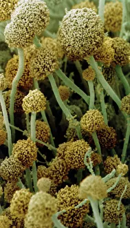
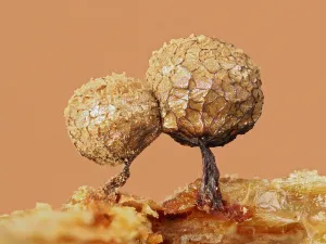
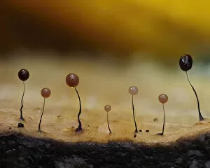
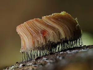
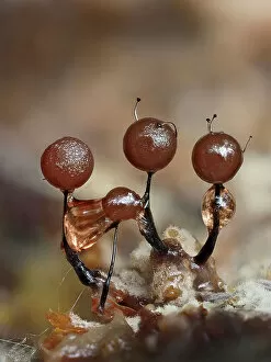
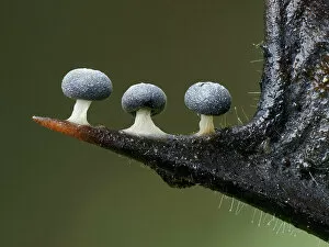
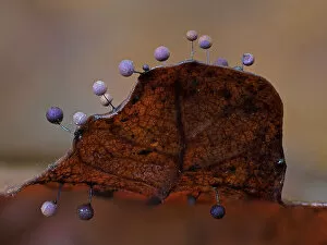
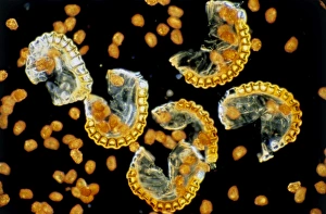
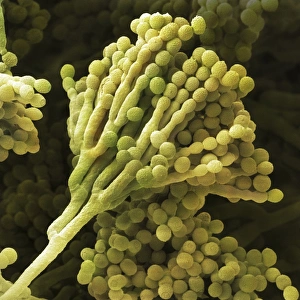
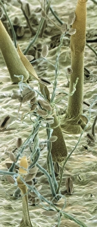
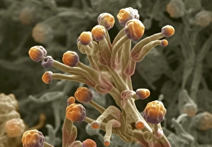
Fruiting bodies of Rhizopus oligosporus, commonly known as sporangium, have been a subject of fascination since their discovery in 1869


All Professionally Made to Order for Quick Shipping
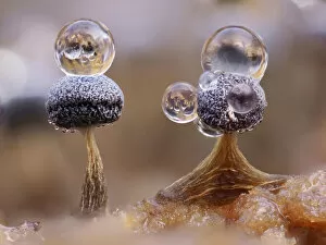
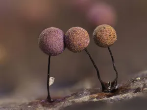
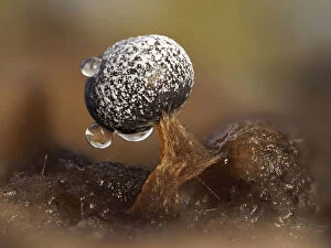
Fruiting bodies of Rhizopus oligosporus, commonly known as sporangium, have been a subject of fascination since their discovery in 1869. These mushroom-like structures are found in bread mould and slime mould species, showcasing the diverse world of fungi. In one captivating image captured by a scanning electron microscope (SEM), we witness the intricate details of bread mould's sporangia. The SEM reveals the complex network of filaments that make up these fruiting bodies, highlighting their role in reproduction. Moving on to slime moulds, we explore the mesmerizing sight of Stemonitopsis typhina sporangia growing along the edge of bark in Buckinghamshire, England. It was November when this stunning display caught our attention with its focus stacked composition. Another striking photograph showcases Metatrichia floriformis' line of split-open sporangia releasing spores. This January moment captured in Buckinghamshire demonstrates nature's ability to create beauty even at microscopic levels. Zooming closer into Lamproderma scintillans' super close-up shot reveals tiny 1mm tall sporangia glistening with life. These small but mighty structures showcase the diversity within slime mould species and remind us that beauty can be found even in the tiniest forms. Dew droplets delicately resting on two Physarum album sporangia provide an enchanting close-up view. This image captures a serene moment where nature meets artistry, reminding us to appreciate life's delicate wonders. Witnessing Stemonitis flavogenita changing from green to orange as it matures on an oak log is truly awe-inspiring. September brings forth this transformational journey right before our eyes through focus-stacked photography techniques used in Hertfordshire, England. August brings about another glimpse into nature's marvels as immature Lamproderma arcyrionema sporangia begin their maturation process.