
Protists Collection
Protists, the microscopic wonders of nature. These incredible organisms are a diverse group that includes protozoans like ciliates and Euglena


All Professionally Made to Order for Quick Shipping
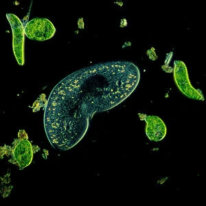
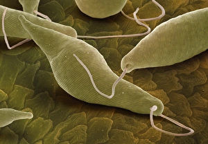
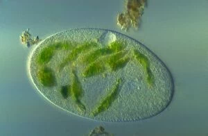
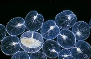
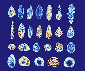
Protists, the microscopic wonders of nature. These incredible organisms are a diverse group that includes protozoans like ciliates and Euglena. In the mesmerizing light micrograph, we can observe a kidney-shaped ciliate surrounded by Euglena sp. , magnified 900 times. The intricate details become apparent, showcasing their unique structures. Plate 14 from Ernst Haeckel's Kunstformen der Natur (Art Forms in Nature) brings us Peridinium Peridinea, captivating our senses with its beauty. This artwork is a testament to the artistic representation and their significance in nature. Euglena gracilis takes center stage in another stunning image captured using scanning electron microscopy (SEM). Its elongated body and flagellum make it an extraordinary specimen worthy of admiration. As we delve deeper into this fascinating world, picture after picture reveals more intriguing aspects of protists. Each snapshot provides a glimpse into their complexity and diversity - Picture No. 11675491, Picture No. 11675490, Picture No. 11675489. . the list goes on. An illustration representing the simplified scheme of evolution showcases how protists play a crucial role as one of three major groups alongside plants and animals, and are integral to understanding the evolutionary history of life on Earth. In yet another light micrograph, we witness the enchanting dance of ciliates under magnification x750 – their graceful movements leaving us awestruck. The final masterpiece comes alive through a powerful light micrograph capturing an elongate ciliate alongside Paramecium bursaria at an astonishing magnification x1500. Their intricately structured bodies reveal hidden worlds within themselves. Protists truly captivate our imagination with their remarkable forms and functions – from elegant shapes to complex behaviors that shape ecosystems worldwide. Exploring these tiny marvels reminds us that even within the unseen realms lies immense beauty waiting to be discovered.