
Duodenal Collection
The duodenum, a vital part of the digestive system, plays a crucial role in the absorption and breakdown of nutrients


All Professionally Made to Order for Quick Shipping
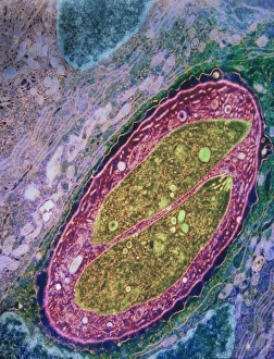
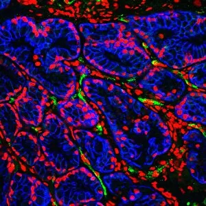
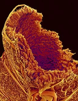
The duodenum, a vital part of the digestive system, plays a crucial role in the absorption and breakdown of nutrients, and is here that the Cholecystokinin-8 molecule (C014 / 4895) and (C014 / 4894) are released, regulating digestion and signaling satiety. However, this intricate organ can also be susceptible to various conditions. Stomach cancer can affect the duodenum, often detected through barium X-ray imaging. Additionally, secondary bowel cancer may manifest in this region as seen in endoscopic view C016 / 8337. Exploring further into the duodenal landscape reveals fascinating structures such as the major duodenal papilla (endoscopic view C016 / 8324), which serves as an entry point for bile and pancreatic secretions. The small intestine's endoscopic view (C016 / 8323) showcases its remarkable absorptive capabilities. Examining closely at the lining of the duodenum (endoscopic view C016 / 8321), we witness its unique composition responsible for nutrient absorption. Furthermore, diverticulum formation within this area is depicted in image C016 / 8320. Microscopic organisms like Isospora sp. Parasites captured via TEM highlight potential infections that can affect this section of our digestive tract twice over with their presence shown by two images consecutively. Delving deeper into microscopic details unveils Brunners glands residing within the duodenum—an essential component secreting mucus-rich fluids aiding digestion. Meanwhile, SEM imagery portrays blood vessels originating from intestinal walls supplying oxygenated blood to sustain proper functioning.