Fraud Blocker Skip to main content

Dividing Collection (page 6)

"Exploring the Intricate Process of Dividing: From Horses to Microorganisms" In a tranquil stable

Background imageDividing Collection: Mouth cancer cell dividing, SEM

Mouth cancer cell dividing, SEM
Mouth cancer dividing. Coloured scanning electron micrograph (SEM) of a squamous cell carcinoma (cancer) cell from a human mouth undergoing mitosis (nuclear division)

Background imageDividing Collection: Dividing liver cancer cell, SEM

Dividing liver cancer cell, SEM
Dividing liver cancer cell. Coloured scanning electron micrograph (SEM) of a hepatocellular carcinoma (HCC) cell undergoing mitosis (nuclear division)

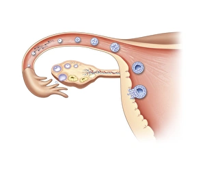
Background imageDividing Collection: Ovulation and fertilisation, artwork

Ovulation and fertilisation, artwork
Ovulation and fertilisation. Computer artwork of the female reproductive system, showing the development of an ovum (egg, small circle) inside an ovary (oval)

Background imageDividing Collection: Dividing cancer cell, SEM C014 / 0361

Dividing cancer cell, SEM C014 / 0361
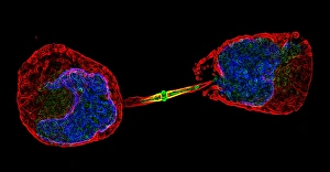
Dividing cancer cell. Coloured scanning electron micrograph (SEM) of a colorectal cancer cell undergoing mitosis (nuclear division) and splitting into two daughter cells (left and right)

Background imageDividing Collection: Dividing brain cancer cells, SEM C014 / 0354

Dividing brain cancer cells, SEM C014 / 0354
Dividing brain cancer cells. Coloured scanning electron micrograph (SEM) of a cancerous astrocyte brain cell that has just undergone cytokinesis (cell division)

Background imageDividing Collection: Dividing bone cancer cell, SEM

Dividing bone cancer cell, SEM
SEM Dividing Osteosarcoma cells Osteosarcoma is an aggressive malignant neoplasm arising from primitive transformed cells of mesenchymal origin (and thus a sarcoma)

Background imageDividing Collection: HeLa cells, SEM C017 / 8305

HeLa cells, SEM C017 / 8305
HeLa cells, coloured scanning electron micrograph (SEM). The cells have just replicated. HeLa cells were the first continuously cultured cell line

Background imageDividing Collection: Lung cancer cell division, artwork C013 / 4630

Lung cancer cell division, artwork C013 / 4630
Lung cancer cell division. Computer artwork of a lung cancer cell during cell division (cytokinesis). The two daughter cells remain temporarily joined by a cytoplasmic bridge (centre)

Background imageDividing Collection: Surfacing lathe

Surfacing lathe with dividing motion and apparatus for cutting wheels. Height of centres 30 inches

Background imageDividing Collection: Young French girl practising her division

Young French girl practising her division
The Four Rules - Division. I am cutting this terrine in two..... but I am eating the whole thing!!! Date: 1905

Background imageDividing Collection: Ciliate protozoa, light micrograph

Ciliate protozoa, light micrograph
Ciliate protozoa. Light micrograph of a mixed population of freshwater ciliate protozoa and algae. The algae are the green filaments crossing the field

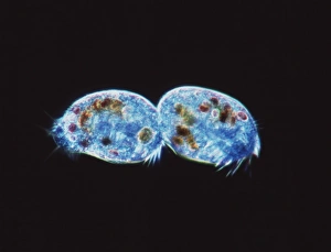
Background imageDividing Collection: Ciliate protozoa dividing

Ciliate protozoa dividing
Ciliate protozoa. Light micrograph of unidentified ciliate protozoa (class Ciliata) undergoing asexual reproduction. These unicellular micro-organisms can multiply by either of two methods

Background imageDividing Collection: Cleavage in frog egg, light micrograph

Cleavage in frog egg, light micrograph The division of cells in an early embryo is called cleavage. These cells will eventually develop into a tadpole and then into a frog

Background imageDividing Collection: Cleavage in frog egg

Cleavage in frog egg. The division of cells in an early embryo is called cleavage. These cells will eventually develop into a tadpole and then into a frog

Background imageDividing Collection: Cleavage in frog eggs

Cleavage in frog eggs. The division of cells in an early embryo is called cleavage. These cells will eventually develop into tadpoles and then into frogs

Background imageDividing Collection: Mitosis, fluorescence micrograph

Mitosis, fluorescence micrograph
Mitosis. Fluorescence micrograph of a cell (centre) in anaphase during mitosis (nuclear division). During mitosis two daughter nuclei are formed from one parent nucleus

Background imageDividing Collection: Embryonic stem cells

Embryonic stem cells. Computer-enhanced confocal light micrograph of dividing stem cells from the ventricular zone of the retina of a developing embryo

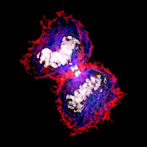
Background imageDividing Collection: Cytokinesis

Cytokinesis. Fluorescent micrograph of an animal cell during cytokinesis (cell division). Cytokinesis occurs after nuclear division (mitosis), which produces two daughter nuclei

Background imageDividing Collection: Abnormal mitosis

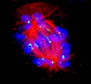
Abnormal mitosis
Mitosis. Fluorescence micrograph of a cell during abnormal anaphase of mitosis (nuclear division). During mitosis two daughter nuclei are formed from one parent nucleus

Background imageDividing Collection: Mitotic cell division, TEM

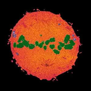
Mitotic cell division, TEM
Mitotic cell division. Coloured transmission electron micrograph (TEM) of a cell during the metaphase stage of mitosis (cell division)

Background imageDividing Collection: Gender determination

Gender determination, computer artwork. Gender is determined by the sex chromosomes, X and Y. Females have 2 X chromosomes and men an X and a Y chromosome

Background imageDividing Collection: Mitosis, TEM

Mitosis, TEM
Mitosis, coloured transmission electron micrograph (TEM). Longitudinal section through a human T cell in metaphase. During mitosis two daughter nuclei are formed from one parent nucleus

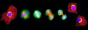
Background imageDividing Collection: Cell mitosis

Cell mitosis. Confocal fluorescence light micrograph composite showing 6 stages of mitotic cell division. At far left, the cell has completed the first stage of cell division known as interphase in

Background imageDividing Collection: Cell division

Cell division. Fluorescent micrograph of an animal cell during cytokinesis (cell division). Cytokinesis occurs after nuclear division (mitosis)

Background imageDividing Collection: Dividing cell, light micrograph

Dividing cell, light micrograph
Cell division. Fluorescent light micrograph of a cell that has divided by mitosis, the asexual replication of a cell into two new cells

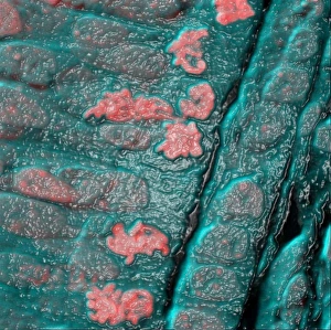
Background imageDividing Collection: Liver cells, SEM

Liver cells, SEM
Liver cells. Scanning electron micrograph (SEM) of liver cells or hepatocytes. Several of the cells (for example, at upper left) are undergoing cell division

Background imageDividing Collection: Technician dissecting a human brain

Technician dissecting a human brain. A technician slices a human brain into 1.5 centimetre thick sections. It has been donated for medical research at the Harvard Brain and Tissue Resource Centre

Background imageDividing Collection: HaCaT culture cell, light micrograph

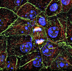
HaCaT culture cell, light micrograph
HaCaT cell. Immunofluorescence light micrograph of a HaCaT cell dividing into two. The nucleus, which contains the cells genetic information, is purple

Background imageDividing Collection: Mitosis, artwork

Mitosis, artwork

Background imageDividing Collection: Dividing bacteria, artwork

Dividing bacteria, artwork

Background imageDividing Collection: Protocell proliferation, artwork

Protocell proliferation, artwork
Protocell proliferation. Image 5 of 5. Artwork showing a protocell (artificial cell) dividing to produce two daughter cells

Background imageDividing Collection: DNA

DNA
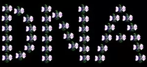
Deoxyribonucleic acid. Conceptual artwork of DNA (deoxyribonucleic acid) spelt out with fluoresence micrographs of anaphase stage cells. Anaphase is a stage of mitosis (nuclear division)


All Professionally Made to Order for Quick Shipping

"Exploring the Intricate Process of Dividing: From Horses to Microorganisms" In a tranquil stable, two horses stand side by side while a small terrier curiously observes their peaceful coexistence. Under the lens of a light micrograph, we witness the mesmerizing beauty of mitosis as cells divide and multiply, creating new life. Delving deeper into the microscopic world, dividing yeast cells come alive in stunning detail through scanning electron microscopy (SEM). The resilient E. Coli bacteria demonstrate their remarkable ability to divide and propagate, showcasing nature's intricate mechanisms on a minuscule scale. Fluorescent micrographs unveil the captivating process of cell division, where vibrant hues highlight each step towards growth and renewal. Sketches depicting St Kilda in Western Hebrides transport us back in time when human settlements were divided by vast landscapes yet connected through shared experiences. Witnessing embryo development 24-36 hours after fertilization reveals how life begins with rapid cell divisions that lay the foundation for future growth and complexity. Whether it is within organisms or across species boundaries, dividing cells remind us of nature's constant drive for renewal and adaptation. Another glimpse into fluorescent micrographs captures the awe-inspiring dance of cell division as it unfolds within different tissues and organisms alike. Stem cells take center stage under scanning electron microscopy (SEM), displaying their potential to differentiate into various specialized cell types—a testament to our body's regenerative capabilities. Even at such minute scales, E. coli bacterium displays its resilience through transmission electron microscopy (TEM), revealing its ability to divide despite challenging conditions—nature's ultimate survivor.