
Cervical Collection
"Cervical: Exploring the Intricacies of the Human Neck and Spine" Delving into the fascinating world anatomy


All Professionally Made to Order for Quick Shipping
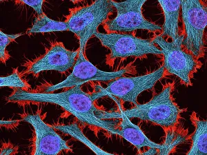
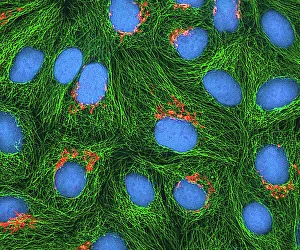
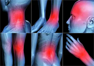
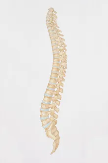
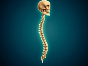
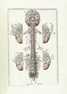
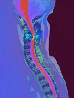
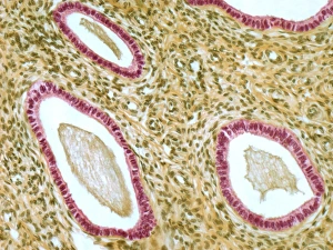
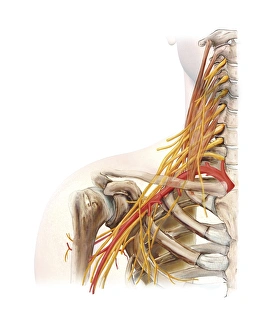
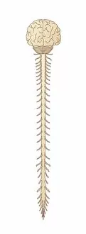
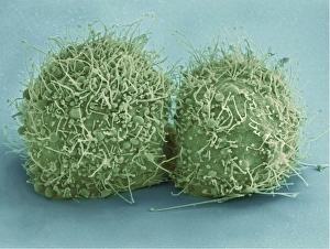
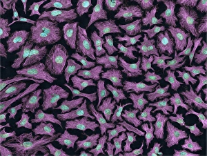
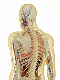
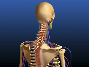
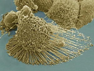
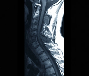
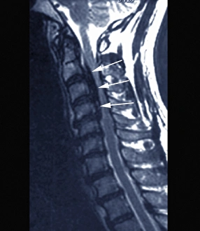
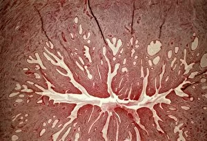
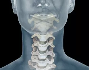
"Cervical: Exploring the Intricacies of the Human Neck and Spine" Delving into the fascinating world anatomy, we encounter HeLa cells under a light micrograph (C017 / 8299), revealing their intricate structure. Another captivating view of HeLa cells (C017 / 8298) showcases their unique characteristics, shedding light on their role in scientific research. Artwork depicting body pain serves as a reminder of the importance of understanding cervical health and seeking proper care when needed. A diagram showcasing the human spine from a side view provides insight into the complexity and interconnectedness of our vertebrae within the cervical region. Through a conceptual image featuring a human skull and spinal cord, we gain an appreciation for how vital this area is to our overall well-being. Bartholomeo Eustachi's "The Science of Human Anatomy" takes us back in time, offering valuable insights into early studies on cervical anatomy that laid foundations for modern knowledge. A coloured X-ray captures a side view of the neck, allowing us to visualize its intricate structures while highlighting any potential abnormalities or injuries. An MRI scan reveals inflamed spinal discs within the cervical region, emphasizing why it is crucial to address such conditions promptly for optimal health outcomes. Captivating artwork dedicated to head and neck anatomy encourages us to explore this complex subject further, appreciating its significance in our daily lives. An artwork from 1844 showcases detailed illustrations spinal nerves—a testament to early anatomists' dedication in unraveling these intricacies long ago. Whether contemplating neck pain or marveling at scientific advancements like HeLa cells or MRI scans, exploring all facets related to "cervical" allows us to appreciate both its beauty and importance in maintaining our overall well-being.