Home > Mary Evans Prints Online > Micro Photography
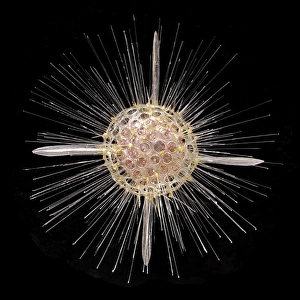
Foraminifer

![]()
Wall Art and Photo Gifts from Mary Evans Picture Library
Foraminifer
Scanning electron microscope (SEM) image of a foraminifer - a single celled organism
Mary Evans Picture Library makes available wonderful images created for people to enjoy over the centuries
Media ID 8595843
© Mary Evans Picture Library 2015 - https://copyrighthub.org/s0/hub1/creation/maryevans/MaryEvansPictureID/10705247
Amoeba Amoebidae Amoebozoa Electron Micrograph Enlarged Eukaryote Eukaryotic Foram Foraminifera Foraminiferan Magnified Micrograph Microscope Image Protist Protista Protozoa Protozoan Retaria Rhizaria Scanning Electron Micrograph Scanning Electron Microscope Scanning Electron Microscope Image Sem Image Tubulinea Tubulinida
EDITORS COMMENTS
1. Title: Magnificent Microcosm: A Scanning Electron Microscope Reveals the Intricate Structure of a Foraminiferan 2.. Delve into the microscopic world and marvel at the intricate beauty of a Foraminiferan, a single-celled organism captured in stunning detail by this Scanning Electron Microscope (SEM) image from Mary Evans Prints Online. Foraminiferans, also known as Foraminifera, are protists belonging to the phylum Rhizaria, with the subclass Foraminifera, and the class Amoebozoa. These eukaryotes, characterized by their complex, tubular tests, are commonly found in aquatic environments. 3. Description: In this image, the Foraminifer's intricate, tubular structure is on full display. The organism's test, a protective shell made of calcium carbonate, is intricately adorned with numerous tiny projections, giving it a textured, almost labyrinthine appearance. The test's complex structure not only provides protection but also plays a role in the organism's feeding and respiration. 4. Magnification and Technique: The image was captured using a Scanning Electron Microscope, a powerful tool that allows for high-resolution, three-dimensional imaging of surfaces. In this case, the Foraminiferan was meticulously prepared and coated with a thin layer of conductive material to enable the electron beam to interact effectively with the sample. The resulting micrograph offers an unprecedented glimpse into the microscopic world, revealing the intricate details of this fascinating organism. 5. Significance: This image serves as a testament to the wonders of the microscopic world and the remarkable capabilities of advanced imaging techniques like the Scanning Electron Microscope. By providing a detailed look at the complex structure of a Foraminiferan, it not only sheds light on the biology of these organisms but also highlights the importance of continued research into the diverse and intriguing world of protists.
MADE IN THE USA
Safe Shipping with 30 Day Money Back Guarantee
FREE PERSONALISATION*
We are proud to offer a range of customisation features including Personalised Captions, Color Filters and Picture Zoom Tools
FREE COLORIZATION SERVICE
You can choose advanced AI Colorization for this picture at no extra charge!
SECURE PAYMENTS
We happily accept a wide range of payment options so you can pay for the things you need in the way that is most convenient for you
* Options may vary by product and licensing agreement. Zoomed Pictures can be adjusted in the Cart.